7.1 POJA-L1921+1919+1920
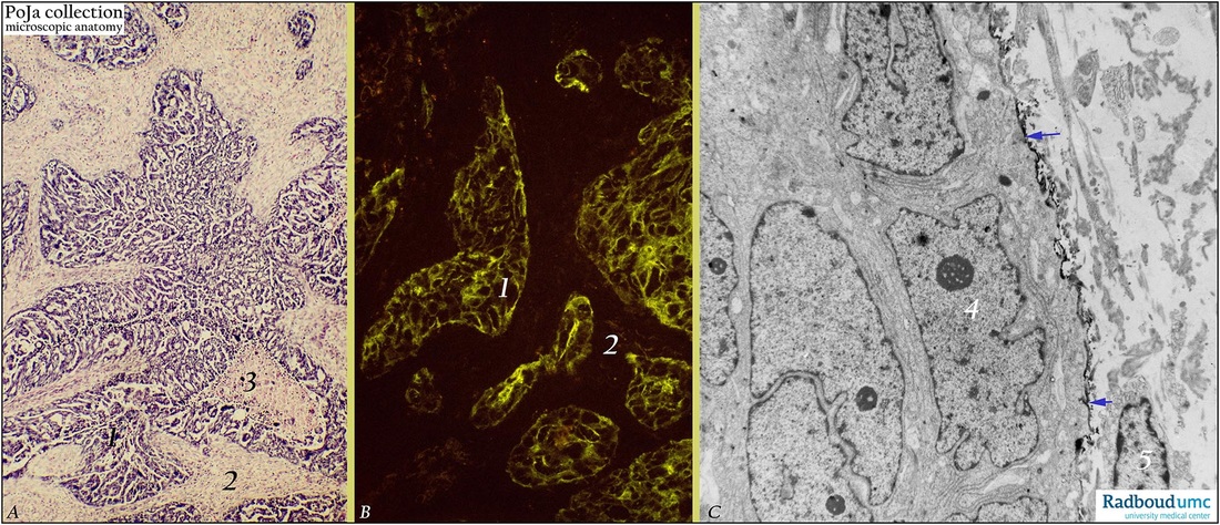
Title: Papillary serous cystadenocarcinoma of ovary (human, adult)
Description: Case nr. p877. Stain: (A) Hematoxylin-eosin; (B) Fluorescent isothiocyanate (=FITC)-labeled OC125 antibody (immunofluorescence, frozen section); (C) immunoelectron microscopy using peroxidase with diaminobenzidin reaction (DAB) OC125 antibody.
(A): Survey of well-differentiated papillary serous cystadenocarcinoma. Packed papillary invasion consists of extended tubuloacinar formation (1). Desmoplastic stroma (2) and necrotic area of tumor cells (3)
(B): Not all tumor cells (1) in cross-sectioned tubuloacini react homogeneously strong with OC125. Intensity of linear fluorescence along tumor cell membranes varies between individual cells and some cells appear negative. Stroma shows no fluorescence (2).
(C): Ultrastructure of an epithelial islet of tumor cells with large nuclei (4) and distinct nucleoli. Specific membrane-bound OC125 reactivity is observed as an electron-dense product only at the free surface of peripheral cells (arrows). No reaction product is present in the close apposed inner cells. Fibroblast (5) and extensions and collagen in stroma are also devoid of reaction product.
Background: See also Background POJA-L1476+L1760+L1770+L1750 Solid areas of the tumor are composed of closely packed papillae. Destructive infiltrative stromal invasion is evident as the tumor ingrowth is diffuse or as sheets of solid tubuli. The OC125 (MUC16) antibody recognizes an epitope on a molecule called Cancer Antigen 125 (CA125) a.o. in tumor cell membranes. It is directed against a glycoprotein produced by human epithelial ovarian cancer cells. However not all tumor cells react uniformly. The antigenic determinant CA125 is expressed on more than 80% of all non-mucinous epithelial ovarian tumors.
Keywords/Mesh: female reproductive organs, OC125 antibody,, ovarian cystadenocarcinoma, female genitalia, ovarian neoplasms, papillary serous cystadenocarcinoma, immunofluorescence, immuno electron microscopy, histology, POJA collection

