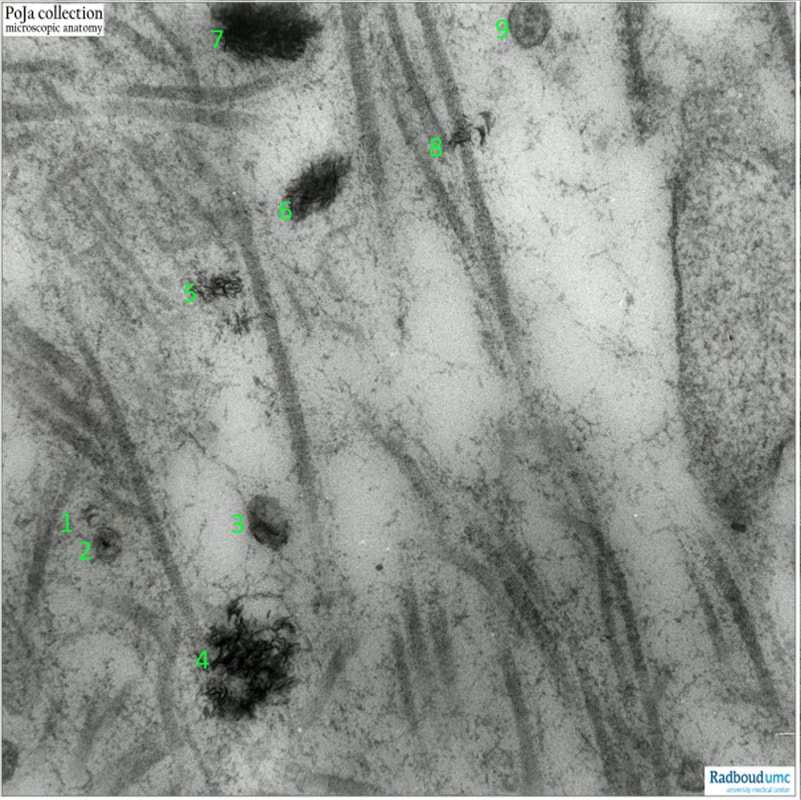
16.1.3 POJA-L7024 Calcification process in bone 2
16.1.3 POJA-L7024 Calcification process in bone 2
Title: Calcification process in bone 2
Description:
Electron micrograph of deposition of calcification crystals (CC) in the production of bone matrix in rat foetus, shown at variable stages and sizes.
(1): Vesicle with one CC,
(2): Vesicle with cross section CC,
(3): Larger vesicle with CC,
(4): Larger amount of CC,
(5 and 8): CC present without detectable vesicles,
(6 and 7): Large amount of CC, however without a visible vesicle,
(9): Small vesicle with proteinaceous dots, but without CC's.
Background
Note the network of collagen fibrils in the matrix.
The mineralised collagen fibrils are formed by the combination of collagen fibrils and hydroxyapatite (HA) mineral crystals. The crystals appear in the form of platelets, approximately 3 x25 x 50 nm in size, although significant variations in platelet size have been reported. The crystals are either intra- or extra-fibrillar, where intrafibrillar crystals are associated with the gap regions of the collagen fibril, while extra-fibrillar crystals are found in the space surrounding the fibrils. It is worth noting that the collagen–mineral interaction is a topic of intense interest and study.
Keywords/Mesh: bone, foetus, electron microscopy, calcification crystals, matrix, POJA collection

