1.1 POJA-L649
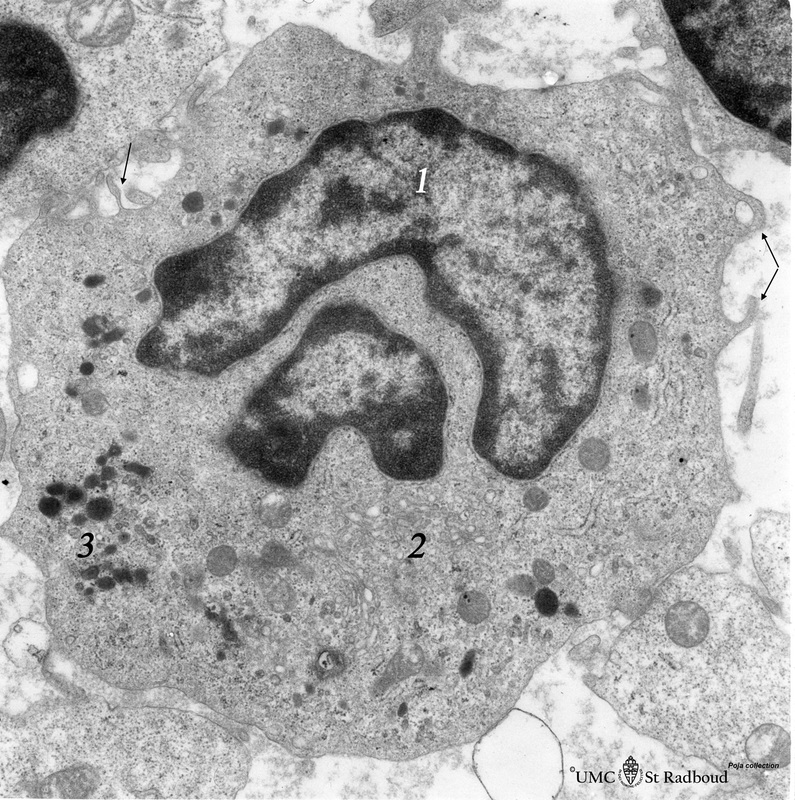
Title: Monocyte (peripheral blood, human)
Description: Electron microscopy.
A large cell with an indented nucleus (1), few Golgi areas (2) and numerous organelles. The cytoplasm contains scattered homogeneous electron-dense lysosomal granules (3) (azurophilic granules with acid phosphatase, arylsulfatase) in variable amounts and few vacuoles. Small pseudopodia (↓, arrows) reflect the ameboid movement and phagocytic ability.
Background: The monocytes (2-8%) of the total leukocyte population are the largest leukocytes (12-20 mm). They travel briefly in the bloodstream (20 hours) and enter thereafter in peripheral tissues as macrophages. They can transform as well into antigen-presenting cells (APC’s).
Keywords/Mesh: blood, bone marrow, monocyte, lysosome, phagocytosis, histology, electron microscopy, POJA collection

