7.1 POJA-L1560+1575
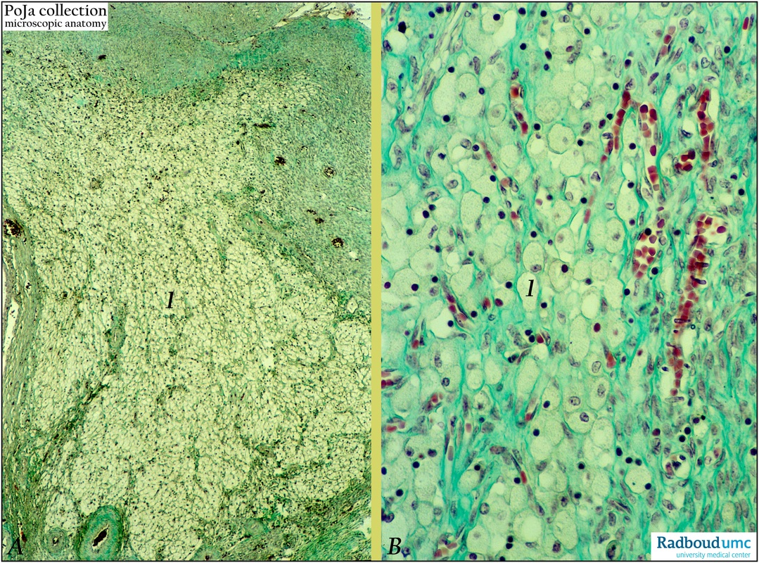
Title: Corpus luteum of menstruation in ovary (human)
Description: Stain: (A, B) Trichrome (Goldner).
(A): Low magnification a degenerating corpus luteum of menstruation (spurium, cyclicum). Epithelioid arranged white lutein cells (1) with abundant lipid show many pyknotic nuclei. Irregular septa and strands of green-stained connective tissue present between lutein cells.
(B): Higher magnification shows the majority of granulosa lutein cells (1) with foamy cytoplasm (lipid). Numerous dark-stained pyknotic nuclei, fibrous connective strands and thin-walled blood vessels are remarkable.
Background: After ovulation the lining stratum granulosum of the empty follicle collapses, becomes folded. Under influence of luteinizing hormone (LH) the luteinization process starts i.e. the granulosa cells increase in size and differentiate into granulosa lutein cells while they produce progesterone/estrogen as well as a yellow carotenoid pigment (lutein). Hence the name corpus luteum due to the yellowish color. Internal theca cells invade in the cellular mass together with capillaries and connective tissue and they differentiate into theca lutein cells (or paralutein cells). Together with the granulosa lutein cells, they form epithelial-like clusters and sheets of round or polygonal lutein cells with fat droplets all together constituting the parenchyma of the corpus luteum. A corpus luteum of menstruation resembles the well-known large, bright yellow corpus luteum of pregnancy; however, the former is smaller and has a more orange-yellow color. Additionally many theca interna and externa cells involute and appear as small spindle-shaped cells. A corpus luteum of menstruation (corpus luteum cyclicum, spurium) may exist for about 2 weeks if no fertilization of the ovum takes place. After luteolysis (involution of corpus luteum) a cascade of apoptosis, fatty degeneration of lutein cells, autolysis and removal by macrophages follows. Gradually a small fibrous hyaline-like scar of acellular collagenous tissue is left after about two months and embedded within fibrous ovarian stroma.h
Keywords/Mesh: female reproductive organs, ovary, menstruation, corpus luteum, luteal cells, female genitalia, histology, POJA collection

