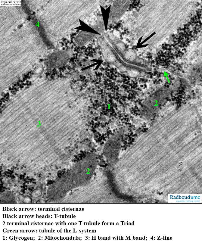
14.1 POJA-L6248 Electron micrograph of the triad structure in striated muscle (human)
14.1 POJA-L6248 Electron micrograph of the triad structure in striated muscle (human)
(By courtesy of L. Eshuis BSc Section Electron microscopy, Department of Pathology, Radboud university medical center, Nijmegen, The Netherlands)
Title: Electron micrograph of the triad structure in striated muscle (human)
Description:
The image shows the transverse tubule system or T-tube invagination of the sarcolemma deep into the sarcomere at the level of A-I band junctions. The tubule (arrow heads) is closely associated with the swollen terminal cisterns (arrows) of the sarcoplasmic reticulum and serves to propagate the electrical triggering to the myofilaments. The cisterns are specialised longitudinal sarcoplasmic reticulum, the so-called L-system (note the tiny tubule at the green arrow). Between each cistern and the tubule fine dots are detectable.
These dots represent the so-called junctional feet or spikes and they might provide a physical link between the gated Ca++release channel (RyR1 or ryanodine receptors) located within the membrane of the cistern and the voltage sensor proteins (DHPR or dihydropyridine receptor) in the membrane of the T-tubule. Also, the protein junctophilin physically docks the SER cisternae and the T-tubule. When the sarcolemma depolarises, the voltage sensor proteins in the T-tubule membrane are activated. Eventually gated Ca++ release channels in the terminal cistern membranes open resulting in a rapid release of Ca++ into the cytoplasm. Simultaneously Ca++ is transported back into the terminal cisterns by Ca++- activated ATPases in the L-system membrane.
See also:
Keywords/Mesh: locomotor system, skeletal muscle, striated muscle, myofibre, Z-line, sarcoplasmic reticulum (SER), T-tubule, triad, gated calcium-release channel, ryanodine receptor, voltage sensor protein, electron microscopy, POJA collection
(By courtesy of L. Eshuis BSc Section Electron microscopy, Department of Pathology, Radboud university medical center, Nijmegen, The Netherlands)
Title: Electron micrograph of the triad structure in striated muscle (human)
Description:
The image shows the transverse tubule system or T-tube invagination of the sarcolemma deep into the sarcomere at the level of A-I band junctions. The tubule (arrow heads) is closely associated with the swollen terminal cisterns (arrows) of the sarcoplasmic reticulum and serves to propagate the electrical triggering to the myofilaments. The cisterns are specialised longitudinal sarcoplasmic reticulum, the so-called L-system (note the tiny tubule at the green arrow). Between each cistern and the tubule fine dots are detectable.
These dots represent the so-called junctional feet or spikes and they might provide a physical link between the gated Ca++release channel (RyR1 or ryanodine receptors) located within the membrane of the cistern and the voltage sensor proteins (DHPR or dihydropyridine receptor) in the membrane of the T-tubule. Also, the protein junctophilin physically docks the SER cisternae and the T-tubule. When the sarcolemma depolarises, the voltage sensor proteins in the T-tubule membrane are activated. Eventually gated Ca++ release channels in the terminal cistern membranes open resulting in a rapid release of Ca++ into the cytoplasm. Simultaneously Ca++ is transported back into the terminal cisterns by Ca++- activated ATPases in the L-system membrane.
See also:
- 14.1 POJA-L6296-B Scheme excitation-contraction coupling at the Triad junction
- 14.1 POJA-L6057+6059+6060+6061 T-tubules and triads in striated muscle
- 14.1 POJA-L6058+6259+6145B Triad system in skeletal muscle
Keywords/Mesh: locomotor system, skeletal muscle, striated muscle, myofibre, Z-line, sarcoplasmic reticulum (SER), T-tubule, triad, gated calcium-release channel, ryanodine receptor, voltage sensor protein, electron microscopy, POJA collection

