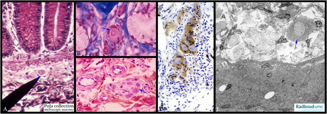
4.1.1 POJA-L3936+La0161+L3937+3938+3939
Title: Nervous plexus of Meissner in intestine (human, guinea pig, rat)
Description: Stain: (A) Hematoxylin-eosin-Heidenhein, colon human. (B) Azan, duodenum, human. (C) Hematoxylin-eosin, colon, human. (D) AMPase, duodenum, guinea pig. (E) Electron micrograph, jejunum, rat.
The plexus of Meissner (arrows, ↘↘) is located underneath the lamina muscularis mucosae (A, 5), i.e. in the loose connective tissue of the submucosa layer. The plexus comprises ganglion cells surrounded by small mantle layer cells (2) (glia-like cells) and delicate nerve fibers towards muscle cells. The mantle layer cells are strongly stained positively for AMPase (D).
In (A, B) the crypts (3) are visible. This internal or intramural nervous plexus and its sympathetic and parasympathetic branches affect both the movement of muscular layers and the secretion activity of epithelial lining and corresponding glands.
In (E, 4) the layer of internal circular smooth muscle cells is shown.
Keywords/Mesh: small intestine, Plexus of Meissner, ganglion cells, electron microscopy, histology, POJA collection

