
2.1 POJA-L911. Hassall’s corpuscle in thymus (mouse)
2.1 POJA-L911
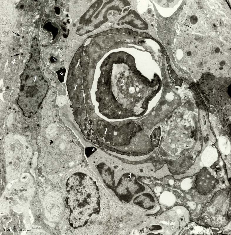
Title: Hassall’s corpuscle in thymus (mouse)
Description: Electron microscopy.
The centre of a small Hassall’s corpuscle consists of darker-stained cells (1) which are keratinizing and represent degenerating cytoplasmic remnants (2).
(3): Is an infiltrating monocyte and (*) indicate the presence of free keratohyalin granules from disintegrated epithelial cells.
(4): Flattened specialized epithelioreticular cells are closely apposing to each other and show desmosomes (→).
Keywords/Mesh: lymphatic tissue, thymus, Hassall’s corpuscle, thymic corpuscle, histology, electron microscopy, POJA collection