
9.5 POJA-L2871+2888+2890
+La0236.
Medullary zone in the adrenal gland
9.5 POJA-L2871+2888+2890+La0236
Title: Medullary zone in the adrenal gland
Description:
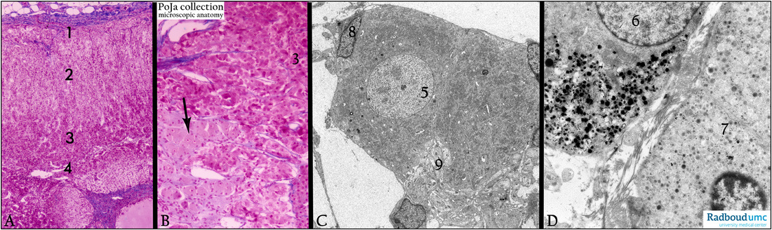
(A, B): Adrenal gland, stain Azan, human< -- >(C, D): Medulla , electron microscopy, rat.
The survey in (A) shows the layers of the cortex and medulla of the adrenal gland. (1) Zona glomerulosa covered by a thin capsule.
(2) Zona fasciculata. (3, 4) Zona reticularis. (B, 3) Catecholamine producing cells in the medulla. The ganglion cells in (B, arrow) and (C, 5) are rich in RER and ribosomes (Nissl substance). (8) Satellite cell. (C, 9) Accumulation of sympathetic nerve endings near the ganglion cell.
Note that in (D) adrenalin (7) and noradrenalin (6) cells show quite different types of granules.
Keywords/Mesh: adrenal gland, cortex, medulla, ganglion cell, adrenalin, noradrenalin, histology, electron microscopy, POJA collection
Title: Medullary zone in the adrenal gland
Description:
(A, B): Adrenal gland, stain Azan, human< -- >(C, D): Medulla , electron microscopy, rat.
The survey in (A) shows the layers of the cortex and medulla of the adrenal gland. (1) Zona glomerulosa covered by a thin capsule.
(2) Zona fasciculata. (3, 4) Zona reticularis. (B, 3) Catecholamine producing cells in the medulla. The ganglion cells in (B, arrow) and (C, 5) are rich in RER and ribosomes (Nissl substance). (8) Satellite cell. (C, 9) Accumulation of sympathetic nerve endings near the ganglion cell.
Note that in (D) adrenalin (7) and noradrenalin (6) cells show quite different types of granules.
Keywords/Mesh: adrenal gland, cortex, medulla, ganglion cell, adrenalin, noradrenalin, histology, electron microscopy, POJA collection