12.1.4 POJA-L4416+4415+2596+3835+3376
Title: Choroid of the uvea
Description:
The uvea or the pigmented vascularised middle tunic is divided into the choroid, ciliary body and iris.
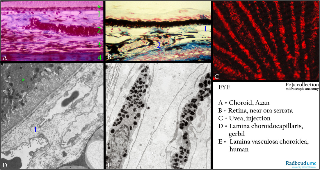
(A): Choroidal stroma, stain Azan, human.
(B): Choroidal stroma, stain Azan, retina near ora serrata, monkey.
(C): Uvea, perfusion in situ with Sirius red, human.
(D): Choriocapillaris, electron micrograph, gerbil.
(E): Choriocapillaris, electron micrograph, human.
The choroid shows three layers (A): Choroidal stroma (2), a connective tissue layer with blood vessels, nerves, few smooth muscles, melanocytes (E). The stroma adheres to the inner pigmented layer (3) (lamina fusca) of the sclera (4).
The choroid is supplied by an extensive network of vessels derived from the long ciliary artery (a branch from the ophthalmic artery).
After in situ perfusion with Sirius red meridional oriented radiating venous branches are demonstrated (C).
Branches of vessels form an intricate network of closely meshed capillaries i.e. the choriocapillaris (1).
Choriocapillaris (A-1, B-1, D-1) with fenestrated capillaries supplies the outer layers of the retina.
The Bruch’s membrane i.e. basement membrane material within a network of elastic and collagen fibres is present in (D-1).
The former is produced by retinal pigmented epithelial cells (*) and endothelial cells of underlying fenestrated capillaries (D).
Background:
The extreme periphery of the retina (B) is known as the ora serrata where an abrupt transition from the inner sensory retina
(pars optica retinae) occurs into the outer nonsensory retina (pars coeca retinae) of the ciliary body and the posterior surface
of the iris. The multilayering of the optically active retina becomes a two-layered epithelium of the pars coeca (optically non-active).
Keywords/Mesh: eye, uvea, choroid, Bruch's membrane, choriocapillaris, retina, ora serrata, histology, electron microscopy, POJA collection

