4.2.1 POJA-La0331+La0268+L3660
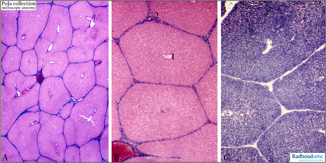
Title: Liver (pig)
Description: Stain: (A, B) Azan. (C) SDH (succinate dehydrogenase.
The liver parenchym cells are radially arranged towards the central vein (A, B, 1) where the blood, flowing through the sinusoids between the liver cells, is collected and transported to ultimately the vena cava inferior.
(2) The portal triade with transverse sections through branches of the arteria hepatica, the portal vein and the bile ductules (C) shows the distribution of the SDH enzyme which is an oxidative marker for mitochondria. In contrast to other rodent species the SDH marker in pig’s liver seems equally distributed through the hepatic lobule (hepaton).
Keywords/Mesh: liver, hepaton, hepatic lobule, SDH, pig, histology, POJA collection

