
12.2.3 POJA-L2601+2605+
2606+2600
Auditory tube in the middle ear
12.2.3 POJA-L2601+2605+2606+2600
Title: Auditory tube in the middle ear
Description:
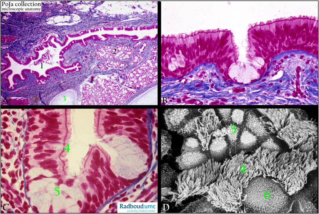
(A - C): Survey lining of auditory tube, stain Azan, human. (A, 1) Eustachian (auditory) tube lined with mutilayered ciliated (respiratory) epithelium with some goblet cells arranged as intraepithelial gland-like structures or invaginations detailed in (B, C, 5).
The middle ear or tympanic cavity is derived from the first pharyngeal pouch lumen. Hence an endodermal-derived epithelium that
extends out from the oral cavity. (A, 2) Seromucous glands around the tuba. (A, green 3) Cartilage.
(D): Scanning electron micrograph of the surface of the ciliated epithelium of the tuba, rat. Kinocilia (compare C, 4 and D, 4).
Gland cells starting to expand (compare C, 5 and D, 5). Large inactive gland cell (D, 6).
Keywords/Mesh: middle ear, auditory tube, eustachian tube, respiratory epithelium, ciliated epithelium, seromucous glands, histology,
POJA collection
Title: Auditory tube in the middle ear
Description:
(A - C): Survey lining of auditory tube, stain Azan, human. (A, 1) Eustachian (auditory) tube lined with mutilayered ciliated (respiratory) epithelium with some goblet cells arranged as intraepithelial gland-like structures or invaginations detailed in (B, C, 5).
The middle ear or tympanic cavity is derived from the first pharyngeal pouch lumen. Hence an endodermal-derived epithelium that
extends out from the oral cavity. (A, 2) Seromucous glands around the tuba. (A, green 3) Cartilage.
(D): Scanning electron micrograph of the surface of the ciliated epithelium of the tuba, rat. Kinocilia (compare C, 4 and D, 4).
Gland cells starting to expand (compare C, 5 and D, 5). Large inactive gland cell (D, 6).
Keywords/Mesh: middle ear, auditory tube, eustachian tube, respiratory epithelium, ciliated epithelium, seromucous glands, histology,
POJA collection