4.1.1 POJA-L4215
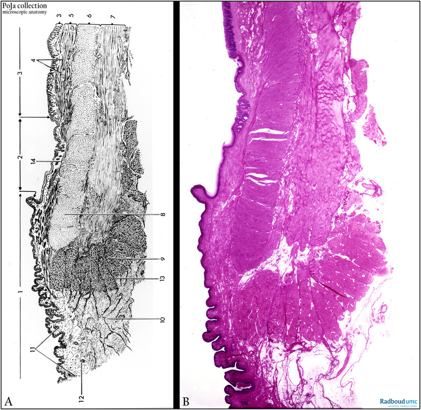
Title: Anal canal or colorectal canal (human)
Description: Stain: (A) Scheme. (B) Hematoxylin-eosin.
The anal canal comprises three zones in the transition from digestive tract into the skin tissue:
(1) Cutaneous zone with keratinizing multilayered squamous epithelium.
(2) Intermediate zone with non-keratinizing multilayered squamous epithelium.
(3) Colorectal zone with simple columnar epithelium forming crypts and containing several distinct epithelial cell types.
(4) Lamina muscularis mucosae.
(5) Tela submucosa.
(6) Inner circular smooth muscle layer.
(7) Outer layer of longitudinal muscle.
(8) Musculus sphincter ani internus, consisting of smooth muscles continuing from (6).
(9) Musculus sphincter ani externus, being striated.
(10) Subcutaneous fat tissue.
(11) Hair follicles.
(12) Apocrine sweat glands called glandulae circumanales.
(13) Septum intermuscularis.
(14) Venous plexus in the tela submucosa (zona haemorrhoidalis).
Background: In contrast to the small intestine the colorectal zone misses the villi. Crypts only are available and the epithelium is rich in goblet cells. The epithelial cells display numerous intercellular interdigitations functioning in water resorption from the stool. Paneth cells are hardly found. The longitudinal muscle layer in the colon is split up in 3 taeniae coli. At the end of the rectum the lamina muscularis mucosae disappears, while the muscularis externa is further specialized into a sphincter ani internus (smooth muscles), followed by a sphincter ani externus that consists, however, of striated muscles. The thin walled venous plexus in this area can give rise to hemorrhoids resulting in fresh blood contamination in the stool.
Keywords/Mesh: colon, rectum, sphincter, scheme, histology, POJA collection

