
7.1 POJA-L1213+1445. Corpus albicans in ovary, post-pregnancy (human)
7.1 POJA-L1213+1445
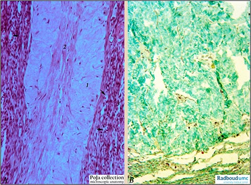
Title: Corpus albicans in ovary, post-pregnancy (human)
Description: Stain: (A) Hematoxylin-eosin (photographed with grey filter); (B) Trichrome (Goldner).
(A): Part of corpus albicans composed of an outer thick fibrous layer (1) and a central part of longitudinal collagenous fibers (2). The scar shows scanty fibroblasts in contrast to the surrounding cell-rich ovarian stroma (3) with many small blood vessels (→).
(B): Part of a similar large corpus albicans composed of relatively acellular collagenous tissue (hyaline-like) which appears characteristically emerald green with Trichrome staining.
Background: There are two types of corpora lutea: (1) By the end of the gestation the diameter of a corpus luteum of pregnancy (verum) (up to 25 mm in the 4th month) is reduced to about 10 mm. Eventually the corpus luteum involutes (luteolysis) and this process involves an apoptotic sequence with the sequential events: (1). Reduction in blood flow within the corpus causes hypoxia. (2). T cells arrive in the corpus and produce interferon which influences the endothelium to attract macrophages. (3). Macrophages produce tumor necrosis factor and then an apoptotic cascade starts. Apoptosis, fatty degeneration of lutein cells, autolysis and removal by macrophages are followed by gradual replacement with fibrous tissue resulting in a large fibrous scar or corpus albicans. This scar will decrease in size but will never disappear. (2) Corpus luteum of menstruation (spurium) (corpus luteum cyclicum) exists about two weeks and after luteolysis a small fibrous scar results. Not all follicles fully develop. Many are lost at various stages of development due to the process of atresia. If atresia occurs at an early stage the follicles e.g. primary follicles completely disappear, but if the process occurs at later stage e.g. secondary follicles the oocyte degenerates and partly due to the collapse of the antrum the zona pellucida and the glassy membrane (membrane vitrea) thicken. Small fibrotic scars (corpora fibrosa or corpora atretica) or even small cysts are the results and can be distinguished by the above-mentioned membranes from corpora albicantia. Gradually all corpora albicantia and corpora atretica (or fibrosa) are pushed towards the medulla and are merged with the ovarian stroma.
Keyword/Mesh: female reproductive organs, ovary, corpus luteum, corpus albicans, female genitalia, histology, POJA collection