
2.2 POJA-L1121. Splenic venous sinus in the red pulp (rat)
2.2 POJA-L1121
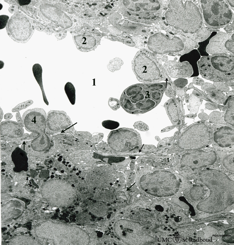
Title: Splenic venous sinus in the red pulp (rat)
Description: Immunoelectron microscopy (gold labelling of heparan sulfate in Lowicryl embedding, using the single chain antibody HS4C3).
(1) Shows the open lumen of a venous sinus filled with few electron-dense erythrocytes and lining cells (2).
(3) Marks a neutrophilic granulocyte. (4) Points to the diapedesis of a lymphocyte through the wall of the sinus.
The sinusoids (cord of Billroth) surround the venous sinus and contain many cells such as lymphocytes and macrophages with dense lysosomes, as well as a few electron-dense erythrocytes.
Arrows indicate the discontinuous basal lamina surrounding the venous sinus as well as the scaffolds of basal lamina material that is reinforced with thin collagen fibrils throughout the splenic cord forming the reticular network of the spleen.
The basal lamina contains electron-dense particles of heparan sulfate molecules (only visible by enlargement of this image). Glycosaminoglycans e.g. heparan sulfate are generally integrated in scaffolds of basal laminae and are secreted by the lining cells and reticular cells.
Keywords/Mesh: lymphatic tissue, spleen, sinus, heparan sulphate, HS4C3 antibody, histology, electron microscopy, POJA collection