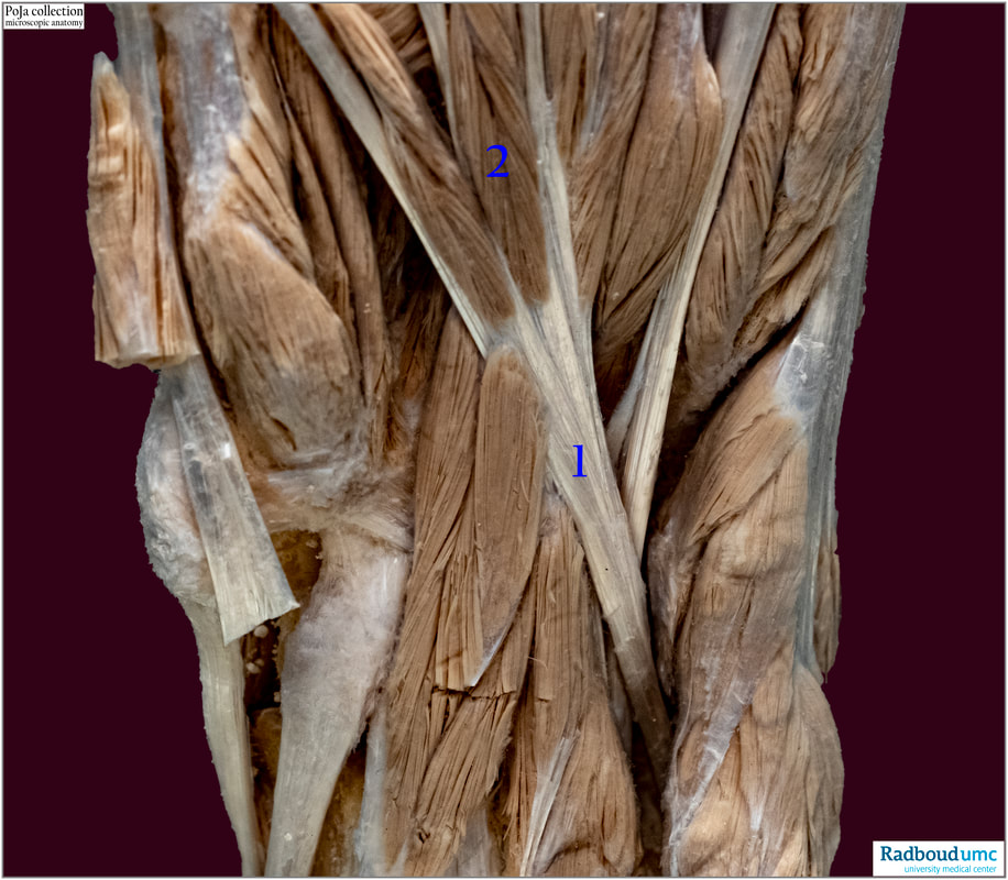
14.5 POJA-L6351 Anatomy of transition of muscles into tendon
POJA-L6351 Anatomy of transition of muscles into tendon
(By courtesy of J. Kooloos PhD and L. Boer PhD, Department Medical Imaging, Anatomy, Museum for Anatomy and Pathology, Radboud university medical center, Nijmegen The Netherlands)
Title: Anatomy of transition of muscles into tendon
Description:
Sole of the foot.
(1): Flexor digitorum longus muscle (sole of the foot).
(2): Lumbricales muscles, which use the tendon as origo.
Note that the tendons are whitish and shining in its attachment into the red-brownish coloured muscle tissue.
Recently fibroblasts have been identified that have switched on a myogenic program. It has been demonstrated that these cells have a dual identity and they fuse into the developing muscle fibres along the musculo-tendon junctions (MTJ) facilitating the introduction of fibroblast-specific transcripts into the elongating myofibres. It is suggested that this mechanism results in a hybrid muscle fibre, primarily along the fibre tips, and enables a smooth transition from muscle fibres characteristics towards tendon features essential for forming robust MTJs.
Fibroblast fusion to the muscle fibre regulates myotendinous junction formation Wesal Yaseen, Ortal Kraft-Sheleg, Shelly Zaffryar-Eilot, Shay Melamed, Chengyi Sun, Douglas P. Millay & Peleg Hasson. Nature Communications volume 12, Article number: 3852 (2021)
See also:
- 16.0 POJA-L7150+7043+7044 Tendon and capsule
- 14.5 POJA-L6078+6079+ 6125 Myotendinous junctions in skeletal muscle
- 14.5 POJA-L6350 Myotendinous junctions in skeletal muscle
Key words/Mesh: locomotor system, muscle, tendon, foot sole, macroscopy, POJA collection

