
13.1 POJA-L4554+4555+4557+4558+4559+4560+4556 Cardiovascular system: light-/electron microscopy of muscle tissues
13.1 POJA-L4554+4555+4557+4558+4559+4560+4556
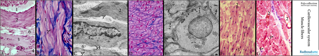
Title: Cardiovascular system: light-/electron microscopy of muscle tissues
Description: In this strip the individual magnifications are irrelevant. The strip shows in a ‘bird’s-eye’ view the muscle tissues.
(A): (POJA-L-4554). Smooth muscle cells in the media of an artery adjacent to an abdominal skeletal muscle, Haematoxylin-eosin, human. Lumen of a cross-sectioned artery (1) with erythrocytes. The wall or media (2) is mainly composed of circular arranged smooth muscle cells (SMC). At (3) longitudinally sectioned and at (4) cross-sectioned striated abdominal muscle cells. (5) Venule and (6) collagen fibres in connective tissue between blood vessel and skeletal muscle. Specific for skeletal muscle cells are syncytia and striation.
(See for more details of skeletal muscle fibres the POJA - collection/Nervous Tissue POJA-L-3278 à 4437 and POJA-L-3294 à3289).
(B): (POJA-L-4555). Longitudinal sectioned smooth muscle cells (wall of deferent duct or ductus deferens): Azan, human. Slender, short fusiform cells (1) with a single central nucleus in a fine fibrillary cytoplasm (=sarcoplasm) surrounded by a blue-stained basal lamina (2, arrow). Note that the SMCs of the deferent duct are shorter and intertwined compared to the SMC of the media of blood vessels (A).
(C): (POJA-L-4557). Electron micrograph of SMCs (digestive tract, gerbil). Central nucleus and organelles i.a. Golgi area (1). Myofilamentous part of sarcoplasm with (2) fusiform densities (or dense bodies). The contractile myofilaments are composed of lattices of thick myosin, thin actin filaments as well as desmin and vimentin. Thin basal lamina (3, arrow) and attachment plaques (4, arrows) (or dense bodies at the sarcolemma). Micropinocytotic vesicles (caveolae) (5, arrow) at the sarcolemma, as well as interstitial collagen fibres (6).
(D): (POJA-L-4556). Cross-sectioned smooth muscle cells (wall of urinary bladder), Azan, human. Each long fusiform smooth muscle cell contains one centrally located nucleus, hence not all cross-sectioned cells exhibit a nucleus. Each cell is wrapped within a thin blue-stained basal lamina intermingled with fine collagenous interstitium. Note that the SMCs are interlaced and packed in bundles.
(E): (POJA-L-4558). Electron micrograph of cross-sectioned smooth muscle cells (urinary bladder, rat). Central nucleus and organelles, contractile myosin and actin filaments. Intermediate filaments desmin and vimentin form with actin and actinin fusiform densities (1) (or dense bodies). Distinct basal lamina (arrow, 2) and (arrow, 3) attachment plaques (dense bodies at the sarcolemma). (Arrows, 4) Pinocytotic vesicles (caveolae) at the sarcolemma.
(F): (POJA-L-4559). Longitudinal sectioned adult cardiocytes of ventricle, Haematoxylin eosin, autopsy human. Note anastomosing pattern (arrow, 1) of the striated cylindrical cells. At (arrow, 2) the intercalated discs. At (3) nuclear poles sometimes filled with lipofuscin. Anastomosing pattern, striation and intercalated discs are specific for cardiomyocytes.
(G): (POJA-L-4560). Cross-sectioned adult cardiocytes, Azan, human. Note characteristic irregular outline of the adult cardiocytes with a diameter up to 1.5 micrometers and a single centrally located nucleus. A thin endomysium surrounds each cell, it consists of a basal lamina ( external lamina) and a thin network of delicate collagenous fibres. (Arrows, 1) Capillaries and (2) venules with a wide lumen and blood cells.
Background:
1. Cardiomyocytes: The striated cells are cylindrical with a single centrally located nucleus. The anastomosing cells are cylindrical and up to 100 micrometers long with a single centrally located nucleus. The cells show dark-stained A-band and light-stained I-bands (c.q. thin Z-lines). Intercalated discs join individual cells. A thin endomysium surrounds each cell, it consists of an external lamina (= basal lamina) and a thin network of delicate collagenous fibres.
2. Smooth muscle cell: These dense bodies are the equivalent of the Z-discs in skeletal and cardiac muscle cells and they may coordinate tensions from the cytoskeleton as well as the contractile system. The intermediate filaments are found interlaced through the cell resembling threads of “fish-net” stockings. During contraction intermediate filaments attached to the dense bands transduce the force to the sarcolemma. Compared to striated muscle cells these SMCs are less organised without a well-organised contractile system of sarcomeres. Contraction results in shortening in all directions and free SMCs move in a corkscrew fashion.
3. Skeletal muscle cell: Myoblasts develop and fuse to a muscle fibre or muscle cell, a long cylindrical multinucleated syncytium surrounded by a thin connective tissue layer the endomysium. A muscle fibre (or cell) contains myofibrils composed of actin and myosin filaments repeated in functional units (sarcomeres) resulting in striation of the fibre. Bundles of muscle fibres are called fascicles that are enclosed by connective tissue (perimysium). Skeletal muscles are under voluntary control in contrast to cardiac muscle and smooth muscle.
Keywords/Mesh: cardiovascular system, vascularisation, blood vessel, smooth muscle, cardiac muscle, skeletal muscle, histology, electron microscopy, POJA collection