
11.2 POJA-L3257+3258.
Myelophagy in a peripheral nerve bundle
11.2 POJA-L3257+3258
Title: Myelophagy in a peripheral nerve bundle
Description:
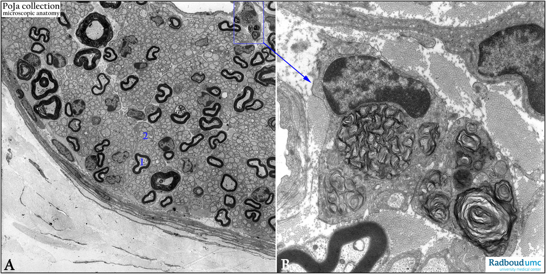
(A, B): Electron micrographs of a peripheral nerve bundle in the principal bronchus of a golden hamster. The nerve bundle in (A) clearly shows the presence of both myelinated nerve fibers (1) and unmyelinated nerve fibers (2). The blue rectangle frame is detailed in (B) and shows myelophagy, i.e. removal of rests of myelin from myelinated nerve fibers by macrophages (myelophages).
Keywords/Mesh: nervous tissue, axon, peripheral nerve fiber, myelinated nerve fiber, unmyelinated nerve fiber, Schwann cell, myelophage, histology, electron microscopy, POJA collection
Title: Myelophagy in a peripheral nerve bundle
Description:
(A, B): Electron micrographs of a peripheral nerve bundle in the principal bronchus of a golden hamster. The nerve bundle in (A) clearly shows the presence of both myelinated nerve fibers (1) and unmyelinated nerve fibers (2). The blue rectangle frame is detailed in (B) and shows myelophagy, i.e. removal of rests of myelin from myelinated nerve fibers by macrophages (myelophages).
Keywords/Mesh: nervous tissue, axon, peripheral nerve fiber, myelinated nerve fiber, unmyelinated nerve fiber, Schwann cell, myelophage, histology, electron microscopy, POJA collection