
12.1.3 POJA-L4414+2558+
2555
Iridocorneal angle of the eye
12.1.3 POJA-L4414 + 2558+2555
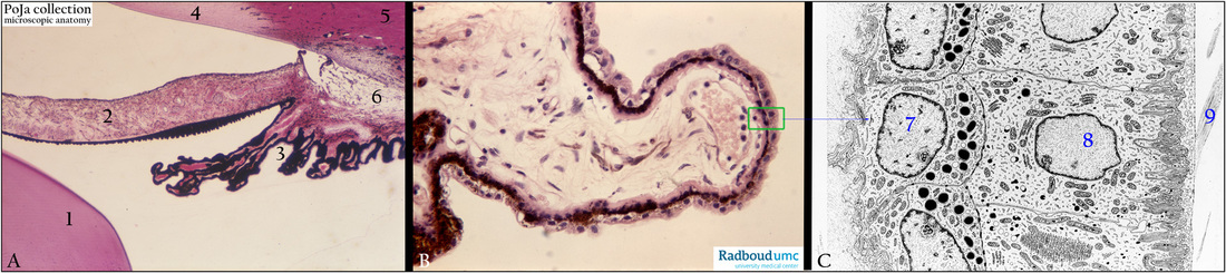
Title: Iridocorneal angle of the eye
Description:
(A): The eye-iridocorneal angle, stain Haematoxylin-eosin, human.
(1) Lens.
(2) Iris.
(3) Ciliary processes.
(4) Cornea.
(5) Limbal stroma.
(6) Corneoscleral trabeculae, loosely arranged sponge-like connective tissue (spaces of Fontana)
continuous with the Schlemm canal.
(B): Ciliary process (pars plicata), stain Haematoxylin-eosin, human.
Detail of the process showing a double epithelial layer of which the inner layer is strongly pigmented.
The green rectangle is schematically described in (C).
From the processes originate short zonular fibres that attach to the lens capsule and enable lens accommodation.
In addition ocular fluid (aqueous humor) is produced by the epithelial cells (supplied by fenestrated capillaries) and
released into the eye chamber.
(C): Electron microscopy scheme of the double epithelial layer of the processes, human. Two cell types are present:
(7) Pigmented ciliary epithelial cells (melanin) with basal infoldings separated by a basal lamina from the ciliary stroma.
(8) Non-pigmented epithelial cells are in intimate apex-to-apex contact with the pigmented ones.
The former (8) also show a basal labyrinth on a basal lamina (inner limiting membrane) separating from the posterior chamber.
The inner limiting membrane increases with age and becomes reticulated.
Within this posterior chamber zonular fibres (9) show numerous fibrils that join the inner limiting membrane and mixed up with the basal reticular netof the non-pigmented epithelial cells (8).
Keywords/Mesh: eye, iridocorneal angle, limbal area, ciliary body , zonular fibre, aqueous humor, histology, electron microscopy,
POJA collection