7.2 POJA-L1555+1556
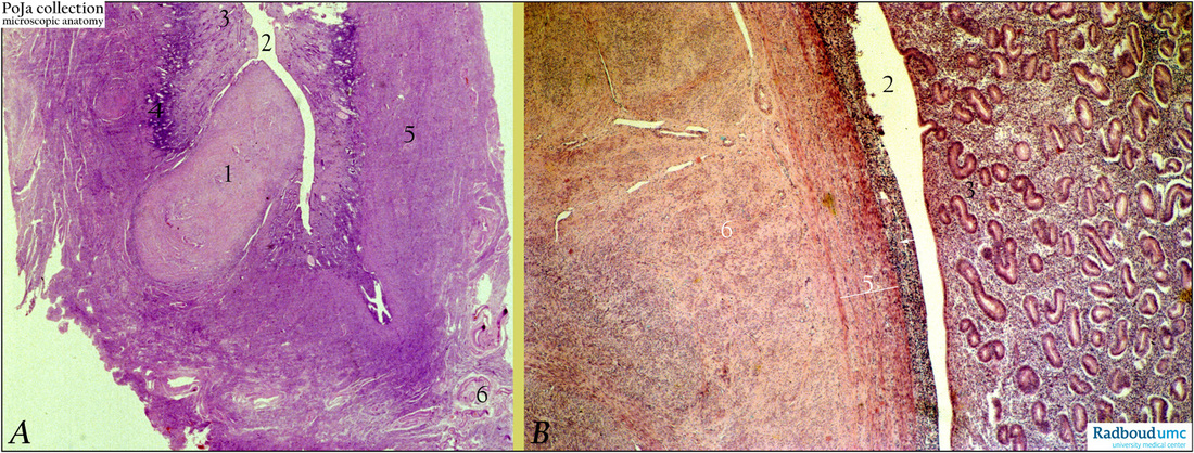
Title: Submucosal leiomyoma (uterus, human)
Description: Stain: Hematoxylin-eosin.
(A): Survey of submucosal leiomyoma (1) bulging into the uterine cavity (2). Endometrium in an early proliferative phase with functional layer (3) and basal layer (4). (5) Middle layer of myometrium and (6) branches of uterine artery (arcuate arteries).
(B): (2) uterine cavity; at (3) another section with endometrial tissue and functional layer (glands in proliferative phase). At the opposite side the lining epithelium (→) is faintly visible and the endometrial stroma is squeezed, partly damaged and infiltrated with lymphocytes due to the proliferation of the myoma. (| 5) is part of the internal layer of the myometrium (stratum submucosum) and at (6) eosinophilic smooth muscle cells of the myoma localized in whirly as well as intertwining bundles. The myoma is well demarcated from normal myometrial smooth muscle cells (5). (Partly by courtesy of G. P. Vooijs MD PhD, former Head of the Department of Pathology, Radboud university medical center, Nijmegen, The Netherlands).
Keywords/Mesh: female reproductive organs, uterus, endometrium, leiomyoma, proliferative phase, smooth muscle cells, histology, POJA collection.

