
16.1.2 POJA-L7155 The anatomy of the Foot
16.1.2 POJA-L7155 The anatomy of the Foot
(By courtesy of J. Kooloos PhD and L. Boer PhD Department Medical Imaging, Anatomy, Museum for Anatomy and Pathology, Radboud university medical center, Nijmegen The Netherlands)
16.1.2 POJA-L7155 The anatomy of the Foot
Title: The anatomy of the Foot
Description:
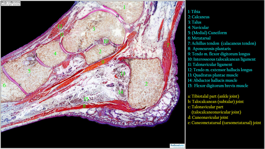
Foot, ankle (human). A modified Mallory-Cason staining procedure for large cryosections. (https://doi.org/10.3109/10520299009105606)
The bones in the foot are formed by endochondral ossification of pre-existing cartilage skeleton. The human foot consists of 26 bones. These bones fall into three groups: the tarsal bones, metatarsal bones, and phalanges. Detailed anatomy of the foot skeleton can be read in the links below (See also:). This sagittal section is meant to illustrate the anatomical relation and orientation between the bones, the ligaments and few muscles.
(1): Tibia
(2): Calcaneus
(3): Talus
(4): Navicular
(5): (Medial) cuneiform
(6): Metatarsal
(7): Achilles tendon (calcaneus tendon)
(8): Aponeurosis plantaris
(9): Tendo. m. flexor digitorum longus
(10): Interosseous talocalcanean ligament (lig talocalcaneum interosseum)
(11): Talonavicular ligament
(12): Tendo m. extensor hallucis longus
(13): Quadratus plantae muscle
(14): Abductor hallucis muscle
(15): Flexor digitorum brevis muscle
(a): Tibiotalal part (ankle joint)
(b): Talocalcanean (subtalar) joint
(c): Talonavicular part (talocalcaneonavicular joint)
(d): Cuneonavicular joint
(e): Cuneometatarsal (tarsometatarsal) joint
The intact epidermis stains reddish for the cornified layer and deep blue for the keratinocytes.
The ligaments, septa as well as tendons are brightly orange-red stained (e.g. 7 and 8) while remnants of muscle bundles are brownish (13-15).
The joints are marked by pink stained cartilage surfaces in (a-e).
See also:
Key words/Mesh: locomotor system, bone, foot, ankle, tibia, tarsal bone, metatarsal bone, ligament, muscle, macroscopy, histology, POJA collection
Title: The anatomy of the Foot
Description:
Foot, ankle (human). A modified Mallory-Cason staining procedure for large cryosections. (https://doi.org/10.3109/10520299009105606)
The bones in the foot are formed by endochondral ossification of pre-existing cartilage skeleton. The human foot consists of 26 bones. These bones fall into three groups: the tarsal bones, metatarsal bones, and phalanges. Detailed anatomy of the foot skeleton can be read in the links below (See also:). This sagittal section is meant to illustrate the anatomical relation and orientation between the bones, the ligaments and few muscles.
(1): Tibia
(2): Calcaneus
(3): Talus
(4): Navicular
(5): (Medial) cuneiform
(6): Metatarsal
(7): Achilles tendon (calcaneus tendon)
(8): Aponeurosis plantaris
(9): Tendo. m. flexor digitorum longus
(10): Interosseous talocalcanean ligament (lig talocalcaneum interosseum)
(11): Talonavicular ligament
(12): Tendo m. extensor hallucis longus
(13): Quadratus plantae muscle
(14): Abductor hallucis muscle
(15): Flexor digitorum brevis muscle
(a): Tibiotalal part (ankle joint)
(b): Talocalcanean (subtalar) joint
(c): Talonavicular part (talocalcaneonavicular joint)
(d): Cuneonavicular joint
(e): Cuneometatarsal (tarsometatarsal) joint
The intact epidermis stains reddish for the cornified layer and deep blue for the keratinocytes.
The ligaments, septa as well as tendons are brightly orange-red stained (e.g. 7 and 8) while remnants of muscle bundles are brownish (13-15).
The joints are marked by pink stained cartilage surfaces in (a-e).
See also:
- 16.1.3 POJA-L7150+7043+7044 Tendon and capsule.
- https://teachmeanatomy.info/lower-limb/bones/bones-of-the-foot-tarsals-metatarsals-and-phalanges/
- https://www.medicalnewstoday.com/articles/324336
- https://www.foothealthfacts.org/articles/when-to-visit-a-foot-ankle-surgeon
Key words/Mesh: locomotor system, bone, foot, ankle, tibia, tarsal bone, metatarsal bone, ligament, muscle, macroscopy, histology, POJA collection