
4.2.1 POJA-L3750+3751+3762. Bile ducts in portal triads of the liver (human)
4.2.1 POJA-L3750+3751+3762
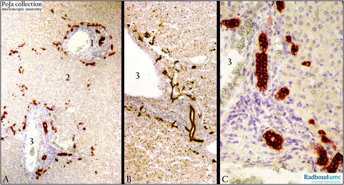
Title: Bile ducts in portal triads of the liver (human)
Description: Stain: Immunoperoxidase staining using antibodies (OVTL12/30) against cytokeratin 7 with DAB (A, B) and AEC (C).
(1) Portal triad with branches of portal vein (3), hepatic artery, brown- or red- stained terminal bile ductules (Hering) and bile ductules.
(2) Liver parenchym cells. The bile ductules are lined by cuboidal cells. The brown- or red- stained clusters of cells between the liver parenchym cells and especially at the periphery of the hepatic lobules are in fact the first adjacent cells of the Hering canals. These terminal ductules form the transition of the liver cells into the bile ductules. The keratin 7 discriminates between liver parenchymal cells and cholangiocytes!!
Keywords/Mesh: liver cells, Hering canal, cytokeratin 7, OVTL12/30 antibody, bile ductule, histology, POJA collection