
12.2.4.1 POJA-L3594+La0132+
3598+3601
Outer hair cells in organ of Corti in the inner ear
12.2.4.1 POJA-L3594+La0132+3598+3601
Title: Outer hair cells in organ of Corti in the inner ear
Description:
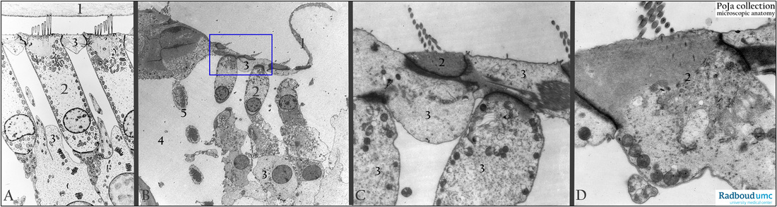
(A): Electron microscopy scheme of part of tectorial membrane (1) and (2) outer hair cells (OHCs), human.
Squeezed between the OHCs phalangeal processes ( cells of Deiter, 3) that contribute to the formation of the reticular membrane (lamina reticularis).
(B): Electron micrograph of three outer hair cells (OHCs) (2), rat. Part of the reticular membrane (1). Phalangeal process (3).
Inner tunnel (4). Outer pillar cell (5).
(C, D): Electron micrographs of hair cells and reticular lamina (or membrane) as outlined in the rectangle in (B), rat.
The phalangeal processes (3) junctioned to the apices of two OHCs (C, 2; D, 2) , form in this section the reticular membrane (lamina reticularis). The stereocilia of the OHCs emerge from a dense actin terminal web in the cell apex (apical cuticular plate).
Note cross-section of central filament in some stereocilia.
Keywords/Mesh: inner ear, organ of Corti, hair cell, stereocilium, lamina reticularis, tectorial membrane, histology,
electron microscopy, POJA collection
Title: Outer hair cells in organ of Corti in the inner ear
Description:
(A): Electron microscopy scheme of part of tectorial membrane (1) and (2) outer hair cells (OHCs), human.
Squeezed between the OHCs phalangeal processes ( cells of Deiter, 3) that contribute to the formation of the reticular membrane (lamina reticularis).
(B): Electron micrograph of three outer hair cells (OHCs) (2), rat. Part of the reticular membrane (1). Phalangeal process (3).
Inner tunnel (4). Outer pillar cell (5).
(C, D): Electron micrographs of hair cells and reticular lamina (or membrane) as outlined in the rectangle in (B), rat.
The phalangeal processes (3) junctioned to the apices of two OHCs (C, 2; D, 2) , form in this section the reticular membrane (lamina reticularis). The stereocilia of the OHCs emerge from a dense actin terminal web in the cell apex (apical cuticular plate).
Note cross-section of central filament in some stereocilia.
Keywords/Mesh: inner ear, organ of Corti, hair cell, stereocilium, lamina reticularis, tectorial membrane, histology,
electron microscopy, POJA collection