
14.1 POJA-L6025+6334+6217 Capillaries, nerves and hypercontraction in skeletal muscles
14.1 POJA-L6025+6334+6217 Capillaries, nerves and hypercontraction in skeletal muscles
14.1 POJA-L6025+6334+6217 Capillaries, nerves and hypercontraction in skeletal muscles
(Fig. B: by courtesy of H. ter Laak PhD Section Neuropathology, retired staff member Department of Pathology, Radboud university medical center, Nijmegen, The Netherlands)
(Fig. B: by courtesy of H. ter Laak PhD Section Neuropathology, retired staff member Department of Pathology, Radboud university medical center, Nijmegen, The Netherlands)
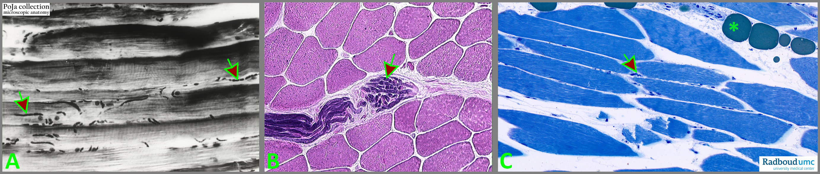
Title: Capillaries, nerves and hypercontraction in skeletal muscles
Description:
(A): Longitudinal running capillaries (arrows) between the muscle fibres in interstitium, shown as dark small tubes (black white print, human).
(B): Bundle of myelinated nerve fibres (arrows) between the muscle bundles, (Sudan black stain, human).
(C): 1 µm plastic section stained with toluidine blue (bovine) showing contraction bands, fat (*) cells and capillaries (arrow). Close to (*) a muscle fibre with spread dense blue spots indicating hypercontraction. Zoom in to see the banding in muscle fibres!
Keywords/Mesh: locomotor system, skeletal muscle, striated muscle, hypercontraction, histology, POJA collection