7.2 POJA-L1579+1546+1682
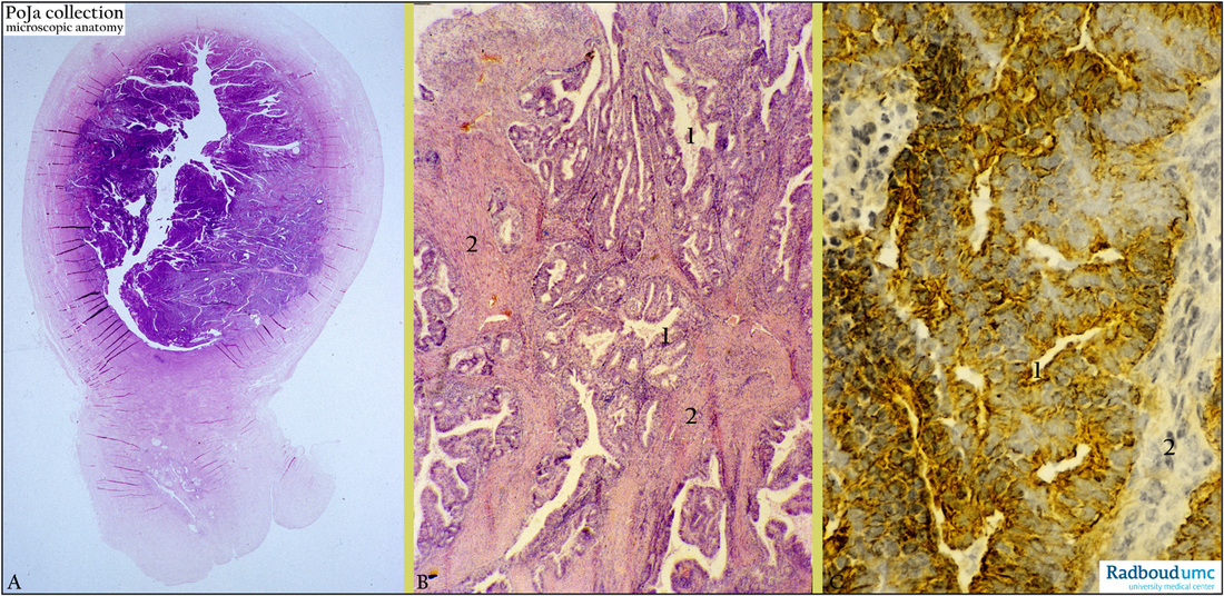
Title: Endometrioid adenocarcinoma, well-differentiated (uterus, human, adult)
Description: Stain: (A, B) Hematoxylin-eosin ; (C) CK 18 antikeratin antibody immunoperoxidase staining with diaminobenzidin reaction (DAB) with hematoxylin counterstaining .
(A): Survey of uterus cavity with exophytic tumor (zoom to observe details). Well-differentiated endometrioid adenocarcinoman with glandular crowding.
(B): Glandular acini (1) on stalks of tight-packed stroma (2) with dispersed inflammatory infiltrations.
(C): Usually cytokeratin 18 (RCK 106) is normally expressed by columnar cells in a.o. urogenital tracts. The tumor give rise to close formation of glands also expressing CK 18 (yellow-brown stain) in lining epithelium (1), stroma (2) remains negative. (Partly by courtesy of G. P. Vooijs MD PhD, former Head of the Department of Pathology, Radboud university medical center, Nijmegen, The Netherlands).
Clinical background: Adenocarcinomas originate from simple lining cells of the endometrium that also forms the glands. The growth pattern of the tumor cells is a reminiscent of normal endometrium. Well-differentiated endometrioid adenocarcinoma is characterised by strong proliferation of endometrial glands. With increasing dedifferentiation the glandular pattern is lost and replaced by papillary formations or solid proliferations. Growth is characterised by friability and spontaneous bleeding, later growth results in myometrial invasion and extending toward the cervix.
Keywords/Mesh: female reproductive organs, uterus, endometrioid cancer, endometrial neoplasms, endometrioid adenocarcinoma, cytokeratin 18, histology, POJA collection.

