
5.4.3 POJA-L3637+2432+
5006+2397+2384+2385.
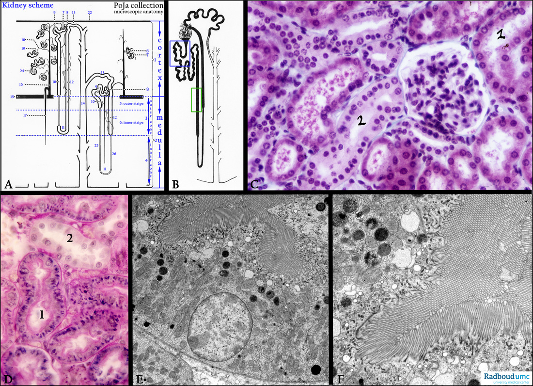
Proximal and distal tubules (I) in the kidney
5.4.3 POJA-L3637+2432+5006+2397+2384+2385
Title: Proximal and distal tubules (I) in the kidney
Description:
(A): Kidney, scheme, human. (Zoom in)
Right side vertical strip with blue characters: (1) Cortex.
(2) Medulla.
(3) Outer zone of the renal medulla.
(4) Inner zone of the renal medulla.
Left side with nephrons:
(1) Cortex.
(2) Medulla, with the outer zone (3) and with the inner zone (4) of the medulla.
(5) Outer stripe of the outer zone.
(6) Inner stripe of the outer zone.
(7) Arteriola efferens leaving the glomerulus.
(8) Macula densa (MD).
(9) Pars contorta I or proximal convoluted tubule (PCT).
(10) Pars recta I or proximal straight tubule (PST).
(11) Henle’s loop (short-looped and long-looped nephrons).
(12) Pars recta II or distal straight tubule (DST) or thick ascending limb (TAL).
(13) Pars contorta II or distal convoluted tubule (DCT); last part is connecting tubule.
(CNT) toward (14) Collecting duct divided in cortical collecting duct (CCD), an outer medullary collecting duct (OMCD) and
an inner medullary collecting duct (IMCD).
(15) Arteria arcuata.
(16) Arteria corticalis radiata (interlobular artery).
(17) Arteriola medullaris recta (vasa recta).
(18) Arteriola afferens.
(19) Arteriola efferens.
(22) Capsula fibrosa.
(24) Glomerulus (glomerular tuft).
(25) Thin ascending limb. (ATL) (long-looped nephron).
(26) Thin descending limb (DTL) (long-looped nephron). Note ATL + DTL = intermediate tubule (IT).
(B): Detailed scheme nephron, human. The blue frame represents the proximal convoluted tubule (PCT), the green frame represents
the proximal straight tubule (PST).
(C): Kidney cortex, stain trichrome Goldner, rat. (1) Proximal convoluted tube PCT, with microvilli. (2) DCT, distal convoluted tubule with hardly any microvilli.
(D): Kidney cortex, stain PAS, mouse. Trypan blue uptake by (re-)absorptive cells of the proximal tubules (1) which are also PAS-positive (brush borders). Distal tubules in cortex (cTAL) lack the microvilli and are much less active in reabsorption.
(E, F): Kidney cortex, electron microscopy, mouse. The proximal tubules show both extended microvilli as well as numerous endocytotic vesicles just below the base of the microvilli. Also note the electron-dense lysosomes in the cells.
Keywords/Mesh: urinary system, kidney, glomerulus, proximal tubule, distal tubule, histology, electron microscopy, POJA collection