
11.5 POJA-L3026+3040+
3027+2969+3366+3039.
Microglial cells and astrocytes in cerebrum
11.5 POJA-L3026+3040+3027+2969+3366+3039
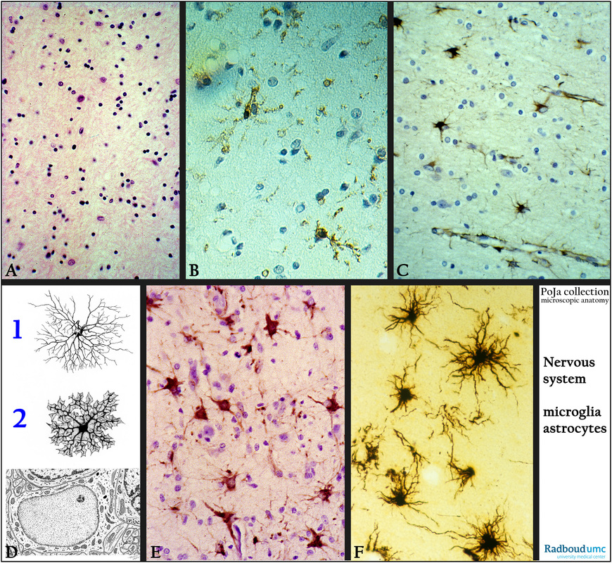
Title: Microglial cells and astrocytes in cerebrum
Description:
(A): Stain hematoxylin-eosin, human. Astroglial cells in the medulla of the cerebrum, the larger nuclei are those of astrocytes and generally
the dense-stained ones are oligodendrocytes. .
(B): Immunoperoxidase staining with DAB and antibodies against HLA-DR, human. Positive microglia cells (antigen-presenting-cells) in cortex, note the spider-like thin branches
(by courtesy of H. ter Laak PhD, Department of Pathology, University Medical Centre Radboud University, Nijmegen, The Netherlands).
(C): Immunoperoxidase staining with DAB and antibodies against GFAP (glial fibrillary acidic protein) an intermediate filament that is
found in glia cells and NOT in ependym), human. The feet or slender processes contact also the capillaries in the cortex.
(D): Electron microscopy scheme of astrocyte, human. Small bundles of intracellular intermediate-sized filaments mostly react with antibodies against glial fibrillary acidic protein (GFAP).
Background: Generally the fibrous asterocyte (F1) reside in the white matter and their processes also form several perivascular endfeet or subpial endfeet.
The protoplasmic asterocytes (F2) are present in the gray matter and at the one side their terminal feet end upon capillaries and are thus involved in the blood-brain-barrier (BBB) by formation of perivascular endfeet (membrana gliae limitans perivascularis). At the other side they end up on neurons.
In addition, several astrocytes create, by their broad subpial endfeet together with a basal lamina (membrana gliae limitans superficialis)
a partition between the central nervous system from the pia mater connective tissue.
(E): Immunoperoxidase staining with DAB and antibodies against GFAP showing the astrocytes in the cortex, human.
(F): Golgi staining of fibrous astrocytes in medulla, note close contact with a dense-stained capillary, human.
(A, C, E, by courtesy of P. Wesseling MD PhD, Head Neuropathology, Department. of Pathology, University Medical Centre
Radboud University, Nijmegen, The Netherlands).
Keywords/Mesh: nervous tissue, cerebrum, oligodendrocyte, microglia, astrocyte, blood-brain-barrier, glial fibrillary
acidic protein, histology, electron microscopy, POJA collection