4.1.1 POJA-L4211
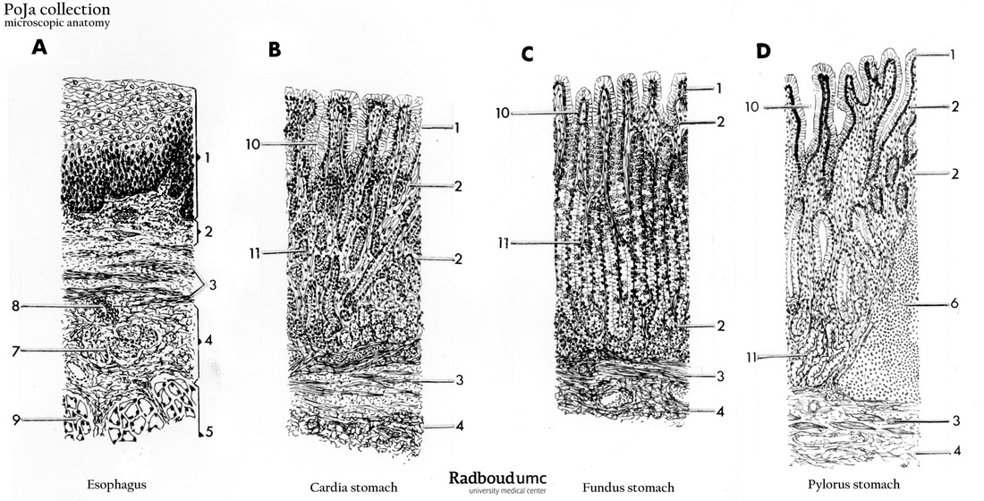
Title: Different parts of esophagus and stomach (human)
Description: Light microscopy scheme:
(A) Esophagus. (B) Cardiac zone of stomach. (C) Fundus zone of stomach. (D) Pylorus zone of stomach.
(1) Epithelium, squamous non-keratinizing in the esophagus, and simple columnar in the stomach zones.
(2) Lamina propria.
(3) Lamina muscularis mucosae.
(4) Tela submucosa (A, B, C).
(5) Tunica muscularis (A).
(6) Foliculus lymphaticus solitaries (D).
(7) Glandulae oesophageae (mucinous) (A).
(8) Glandular duct (A).
(9) Striated muscles (A).
(10) Foveolae gastricae
(B, C, D). Glandulae gastricae (C, 11). Glandulae pyloricae (D, 11). Glandulae cardiacae (B, 11). (C, 6) Lymph follicle.
Background: The function of the stomach is to digest the food entered from the esophagus. The cardiac-zone in the stomach comprises 2-3 cm area at the entrance. The branched and curled glands in the lamina propria produce mucus and some lysozyme and are identical to the esophageal cardiac glands. In the central or corpus or fundus area of the stomach the single layered epithelium creates foveolae (broad short invaginations) which continue further in the straight deep gastric glands. The foveolar epithelial cells produce mucus for protection of their surface. At the transition zone from foveolae to gastric glands the mucous neck cells produce a mucus gel layer for protection. The deeper located large parietal cells produce HCL secreted via a tubulo-vesicular system ending in the intracellular canaliculi of this cell type. The eosinophilic character of the parietal cells is due to the enormous content of mitochondria needed for the HCL production. The production is stimulated by cholinergic nerves, gastrin and histamine. The parietal cell also produces “intrinsic factor” that binds to vitamin B12 thus facilitating the resorption of the complex in the small intestine. At the bottom of the gastric gland most chief cells (basophilic = RER) are located producing pepsinogen and lipases for the digesting of proteins and lipids. Enteroendocrine cells are spread throughout the gastric glands (and elsewhere in the gut) and produce a vast collection of about 30 hormones among which glucagon (A cells), gastrin (G cells), somatostatin (D cells), vasoactive polypeptide (VIP in D` cells), secretin, cholecystokinin, serotonin etc. The enteroendocrine cells are also known as enterochromaffin cells (EEC) or argentaffin cells, or APUD cells (amine precursor uptake). Histologically, the gastric glands look as heterogeneous but straight tubules. The pyloric glands, however, are more branched, curled, and homogenous and mainly produce mucus and lysozyme as well as gastrin. To improve the mobility of the stomach for the food digestion, the tunica muscularis comprises three layers of smooth muscles, the inner diagonal layer, middle circular layer and the outer longitudinal layer.
Keywords/Mesh: esophagus, stomach, cardia, pylorus, fundus, glands, enteroendocrine cells, histology, POJA collection

