4.1.1 POJA-L4142+4147+4146
Title: PAS and CO-TL1 staining of colorectal epithelium (human)
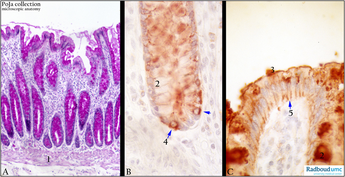
Description: Stain (A) PAS. (B, C) CO-TL1 monoclonal antibody immunoperoxidase staining with (DAB) and hematoxylin counterstaining. The colorectal epithelial lining contains absorptive cells, goblet cells, endocrine cells and stem cells.
(A): Shows that the crypt cells are PAS positive largely due to the goblet cells production of mucins. The most abundant type is MUC2 in colon. (1) Lamina muscularis mucosae.
(B): Shows that also endocrine cells are positively stained with the monoclonal antibody CO-TL1 (4 ↘). The white goblet cells (B, 2) here have released their content, while in (C, 2) they are still present and positive. The colon specific antigen is also transported to both the cell surface (C, 3) and to the lateral cell walls (C, 5).
Keywords/Mesh: colon, PAS, CO-TL1 antibody, mucins, goblet cells, histology, POJA collection

