
12.1.4 POJA-L4422+4423
Fovea centralis versus ora serrata in the retina
12.1.4 POJA-L4422+4423
Title: Fovea centralis versus ora serrata in the retina of the eye
Description:
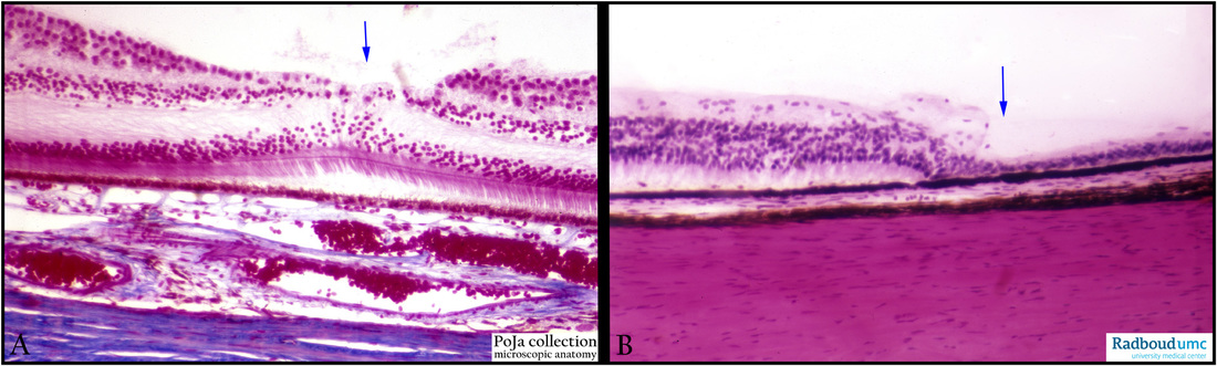
(A): Retina, fovea centralis, stain Azan, human. At the posterior pole of the eye about 4 mm from the optic papilla a yellow spot is called the macula due to local accumulation of xanthophyll (carotenoid) pigments. The central spot in the macula lutea is the fovea centralis with
the foveola as inner centre possessing the sharpest vision since the ganglion cells are moved slightly peripherally in that zone, enabling
the incidence of light directly on the photoreceptor cells.
The retina, here (see arrow), is very thin and mainly contains long, slender cones as well as neuroglial cells (Müller).
The cones synaps ( oblique fibres of outer plexiform layer) with bipolar cells but both are oriented at an angle around the margins of
the foveola.
Locally in the fovea centralis the blood vessels are well developed.
(B): Retina, ora serrata, stain Haematoxylin-eosin, human. The extreme periphery of the retina is the ora serrata because the retinal termination forms serrated processes that extend on the pars plana of the ciliary body. The part of the retina beyond (here right of the arrow) the ora serrata is insensitive for light, simply because the photoreceptor cells and ganglion cells are missing. The outer nuclear layer has reduced into 2-3 layers and gradually nuclei displacement occurs into the outer plexiform layer so that the inner layer and outer nuclear layers are fused. Neuroglia cells replace the absent neural cells. This part is called the pars caeca retinae (nonsensory retina) in contrast to the pars optica retinae (sensory retina). The pigmented layer, however, continues towards the iris.
Keywords/Mesh: eye, retina, macula lutea, fovea centralis, ora serrata, pars caeca retinae, pars optica retinae, histology, POJA collection
Title: Fovea centralis versus ora serrata in the retina of the eye
Description:
(A): Retina, fovea centralis, stain Azan, human. At the posterior pole of the eye about 4 mm from the optic papilla a yellow spot is called the macula due to local accumulation of xanthophyll (carotenoid) pigments. The central spot in the macula lutea is the fovea centralis with
the foveola as inner centre possessing the sharpest vision since the ganglion cells are moved slightly peripherally in that zone, enabling
the incidence of light directly on the photoreceptor cells.
The retina, here (see arrow), is very thin and mainly contains long, slender cones as well as neuroglial cells (Müller).
The cones synaps ( oblique fibres of outer plexiform layer) with bipolar cells but both are oriented at an angle around the margins of
the foveola.
Locally in the fovea centralis the blood vessels are well developed.
(B): Retina, ora serrata, stain Haematoxylin-eosin, human. The extreme periphery of the retina is the ora serrata because the retinal termination forms serrated processes that extend on the pars plana of the ciliary body. The part of the retina beyond (here right of the arrow) the ora serrata is insensitive for light, simply because the photoreceptor cells and ganglion cells are missing. The outer nuclear layer has reduced into 2-3 layers and gradually nuclei displacement occurs into the outer plexiform layer so that the inner layer and outer nuclear layers are fused. Neuroglia cells replace the absent neural cells. This part is called the pars caeca retinae (nonsensory retina) in contrast to the pars optica retinae (sensory retina). The pigmented layer, however, continues towards the iris.
Keywords/Mesh: eye, retina, macula lutea, fovea centralis, ora serrata, pars caeca retinae, pars optica retinae, histology, POJA collection