7.1 POJA-L1509+1510+1905
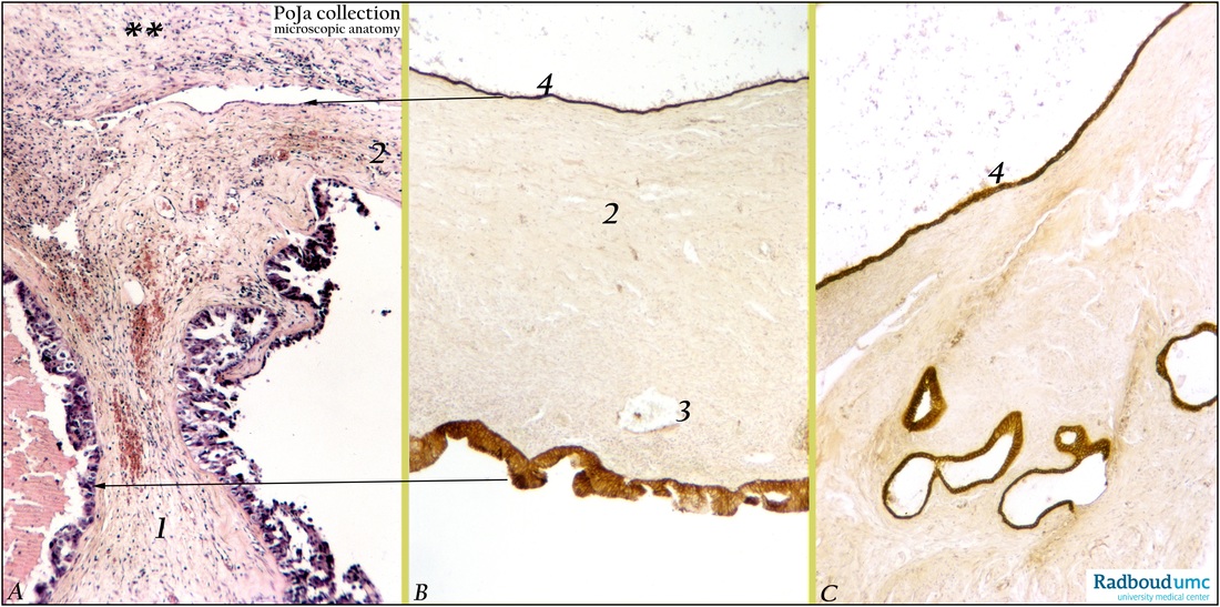
Title: Papillary serous adenocarcinoma of ovary (human, adult)
Description: Stain: (A) Hematoxylin-eosin; (B, C) CK7 (OVTL12/30) antikeratin antibody immunoperoxidase staining with diaminobenzidin reaction (DAB) and hematoxylin counterstaining.
(A): Part of the tumor with two cystic cavities separated by thick septum (1) with blood vessels and local hemorrhages. Fibrous sheet (**) of septum is teared off and partially overlies mesothelial lining (upper arrow point) of capsule (2). Small cavity (left) is crested with bulging tumor cells (lower arrow point); the lumen contains coagulated hemorrhagic fluid. Large cavity (right) shows partly damaged lining of small papillary projections of tumor cells with irregular tufting and nuclear hyperchromatism.
(B): Another section of the same tumor: top side a thick stromal capsule (2) lined by CK7-positive mesothelial cells (4, arrow points to similar mesothelium in A). Capsule with small blood vessels is CK7- negative. Dilated venule (3) contains a metastasized CK7-positive tumor cell closed to lining of large empty cyst (arrow points to similar cystic lining in A). Bulging multilayered tumor cells cover cystic wall and generally show preferably stronger apical staining.
(C): Another section of a similar tumor showing the CK7-positive thin mesothelial lining (4), as well as inclusion cysts regularly lined with epithelial cells.
Background: It is well accepted that normal simple lining epithelia and the epithelium of most mucous or serous glands generally express cytokeratin 7. Ovarian surface epithelial cells are also positive for the low molecular weight keratin protein cytokeratin 7. During abnormal differentiation of these epithelial cells CK7 expression remains preserved. Individual tumor cells of well-differentiated adenocarcinomas metastasize via blood vessels or by invasion. Application of a.o. low-molecular weight keratins such as CK7 allows histological detection of solitary tumor cells in blood vessels more easily and convincingly Histogenetically it is assumed that a majority of epithelial tumors of the ovary is derived from the ovarian serosa. This lining is the equivalent of the embryonic coelomic epithelium in adulthood. Embryologically the coelomic epithelium give rise to the müllerian epithelium (ducts of Müller) and its derivates are the fallopian tube lining (tubal pathway), endometrial lining (endometrial pathway) and endocervical glands lining (endocervical pathway). The ovarian surface epithelium contains undifferentiated cells that possess the latency to differentiate along similar embryonic pathways. In this way a tumor derived from these cells will differentiate analogously. This results in serous tumors formed by epithelial tumors that are differentiated along the tubal pathway. Serous cystadenocarcinomas are essentially a malignant form of serous cystadenomas; they account for ca. 40% of all ovarian cancers and appear to be the most common malignant tumors. The size of malignant serous tumors varies between 10 and 15 cm but sometimes 40 cm diameters are reported. With hemorrhagic foci, the tumors are mostly cystic but also partially solid. The fibrous cysts are lined by tubal-like columnar, often ciliated cells and contain blood-stained serous fluid. Solid areas of the tumor are composed of closely packed papillae that might penetrate the tumor capsule. Luxuriant papillomatous growths might also extend over the surface and obliterate later on the ovarian structure.
Keywords/Mesh: female reproductive organs, ovary, female genitalia, ovarian neoplasms, papillary serous adenocarcinoma, keratin 7, OVTL12/30 antibody, metastasis, mesothelial cells, inclusion cyst, histology, POJA collection

