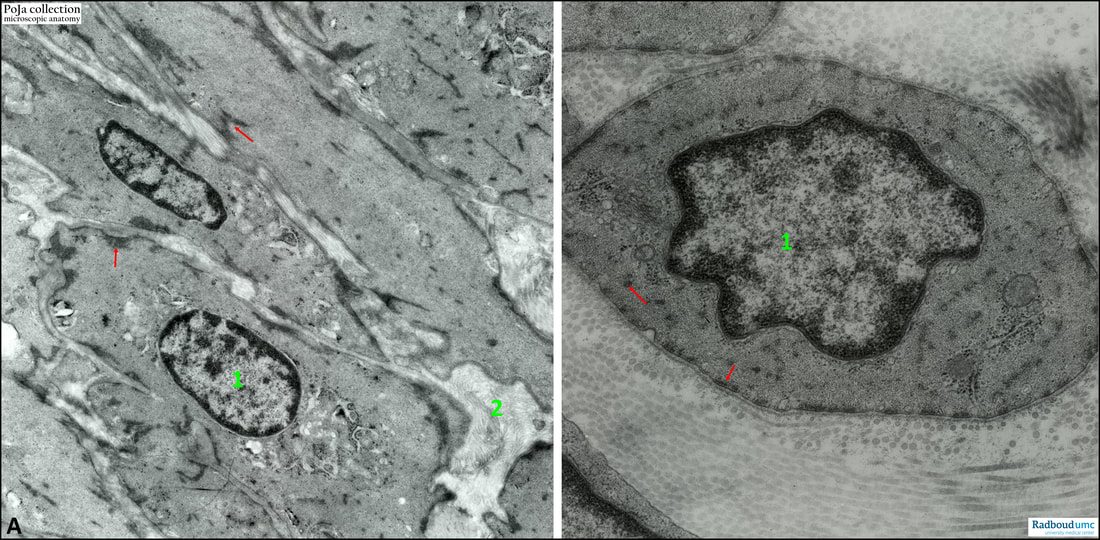
14.3 POJA-L6004+6009 Electron micrograph of smooth muscles of the detrusor in urinary bladder (rat)
14.3 POJA-L6004+6009 Electron micrograph of smooth muscles of the detrusor in urinary bladder (rat)
Title: Electron micrograph of smooth muscles of the detrusor in urinary bladder (rat)
Description:
(A): Longitudinal section and (B): cross section through the smooth muscles of the urinary bladder. Arrows indicate the dense plaques which function as adherence points for the myofilaments. A continuous basal lamina surrounds each individual myocyte (myofibre, muscle fibre cell).
(1): Nucleus of the muscle fibre cell.
(2): Intercellular connective tissue space with collagen fibrils. The detrusor muscle of the urinary bladder functions to filling and emptying the bladder. The muscle fibres are oriented around the bladder and are fibres of variable length.
Keywords: /Mesh: locomotor system, smooth muscle, bladder, dense plaque, electron microscopy, histology, POJA collection

