
12.1.2 POJA-L4397+2538
Stroma of the cornea
12.1.2 POJA-L4397+2538
Title: Stroma of the cornea
Description:
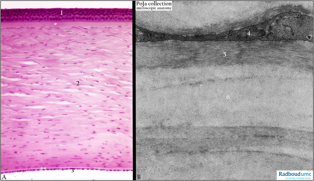
(A): Stain Haematoxylin-eosin, human. At the outside the cornea is covered with 5 to 6 layers of non-keratinising squamous epithelium (1),
settled on the Bowman’s layer (*). At the interior side the cornea is limited by the Descemet’s membrane (**) on which a thin single layer of continuous endothelium (3) is localised. Note that normal corneal stroma is avascular with a relatively low keratocytes density. Due to histological procedures splitting of the collagen bundles occurs.
(B): Electron micrograph of the corneal stroma, human.The thick stroma (A, 2) consists of lamellae of parallel arranged collagen fibrils oriented in two cross directions (5 and 6), and specialised fibroblasts (4) (or keratocytes) in between them.
Keywords/Mesh: eye, cornea, collagen fibril, Bowman’s layer, Descemet’s membrane, histology, electron microscopy, POJA collection
Title: Stroma of the cornea
Description:
(A): Stain Haematoxylin-eosin, human. At the outside the cornea is covered with 5 to 6 layers of non-keratinising squamous epithelium (1),
settled on the Bowman’s layer (*). At the interior side the cornea is limited by the Descemet’s membrane (**) on which a thin single layer of continuous endothelium (3) is localised. Note that normal corneal stroma is avascular with a relatively low keratocytes density. Due to histological procedures splitting of the collagen bundles occurs.
(B): Electron micrograph of the corneal stroma, human.The thick stroma (A, 2) consists of lamellae of parallel arranged collagen fibrils oriented in two cross directions (5 and 6), and specialised fibroblasts (4) (or keratocytes) in between them.
Keywords/Mesh: eye, cornea, collagen fibril, Bowman’s layer, Descemet’s membrane, histology, electron microscopy, POJA collection