
2.2 POJA-L1119. Part of lymphatic nodule in spleen (rat)
2.2 POJA-L1119
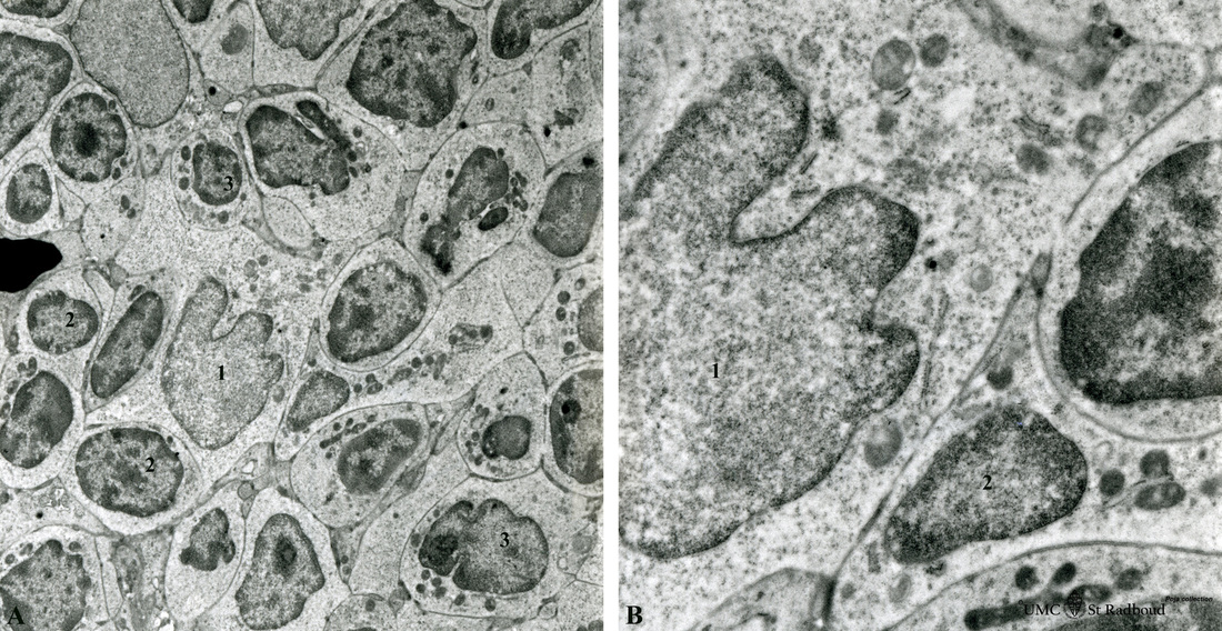
Title: Part of lymphatic nodule in spleen (rat)
Description: Electron microscopy.
The left image (A) reveals part of a white pulp area stuffed with a dendritic cell (1) between a majority of different types of lymphocytes (2, 3). The right image shows a larger magnification of the same area with the dendritic cell (1) sandwiched in between the enclosing lymphocytes (2).
Keywords/Mesh: lymphatic tissue, spleen, white pulp, histology, electron microscopy, POJA collection