
6.2 POJA-L2717+2720+2718.
Ductus epididymides
6.2 POJA-L2717+2720+2718
Title: Ductus epididymis
Description:
(A) Stain Weigert hematoxylin, human. (B, C) Electron microscopy, gerbil.
The epididymal duct is covered with a two-layered pseudostratified columnar epithelium that is androgen-dependent.
The basal cells (A, 1) are small and round and function as replacement cells for the taller columnar principal cells with
an oval nucleus (2). The latter ones are provided with clustered stereocilia (3) detailed in (C, 4).
The lamina propria (A) also contains myofibroblasts. In the ductal lumen different cross-sections of spermatozoa (5) are found.
(B, C) Within the organelles-rich cytoplasm of the principal cells elaborate perinuclear Golgi areas (**) are present for secretion
of a.o. glycoproteins as well as for endocytosis and pinocytosis of the testicular fluid.
Keywords/Mesh: testis, epididymis, epididymal duct, histology, electron microscopy, POJA collection
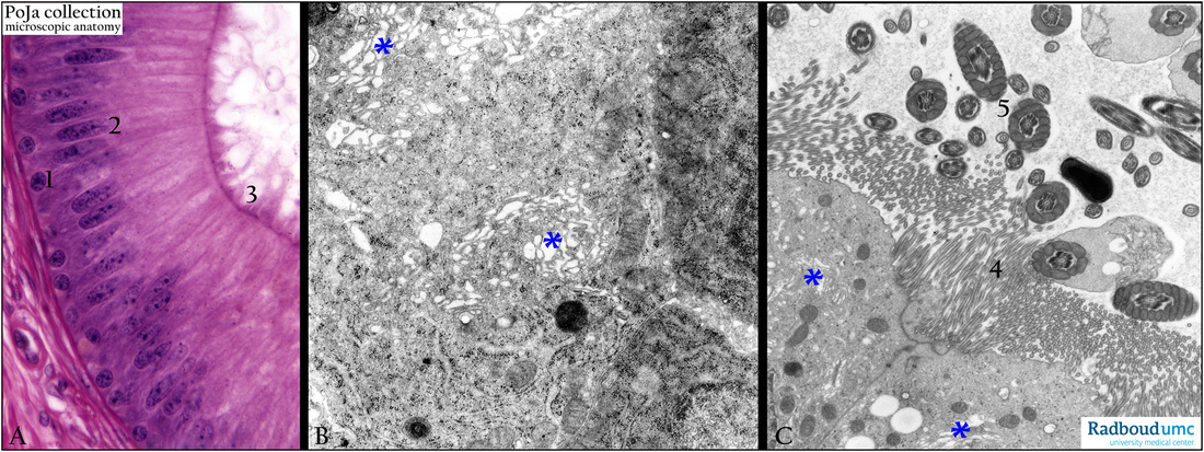