5.4.1 POJA-L2341+2543
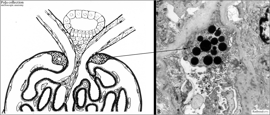
Title: Glomerular periportal cells (XI) in the kidney
Description:
(A): Glomerulus, electron microscopy scheme, toad. (By courtesy of A. Lamers PhD, Department Cell biology and Histology, Radboud university medical center, Nijmegen, The Netherlands).
(B): Glomerulus, electron microscopy, human. Periportal cells are granulated epithelial cells formed around the vascular pole of
the glomerulus. They are present in only a minority of human glomeruli, primarily in outer cortical glomeruli.
These cells form junctional complexes with podocytes and parietal epithelial cells.
The cells are closely associated with renin-containing cells in the afferent arteriole.
They have uniformly electron-dense stained granules which, however, do not contain renin.
Keywords/Mesh: urinary system, kidney, glomerulus, periportal cell, histology, electron microscopy, POJA collection

