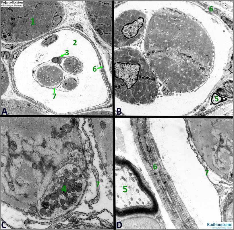
14.1 POJA-L6066+6068+6069+6067 Electron micrograph of muscle spindle (mouse)
14.1 POJA-L6066+6068+6069+6067 Electron micrograph of muscle spindle (mouse)
Title: Electron micrograph of muscle spindle (mouse)
Description:
(A): Striated muscle fibres (1) with peripheral located nuclei. (2): Peraxial space of the muscle spindle (slightly enlarged artificially due to preparation procedures) with external capsule derived from the perimysium (6) and intrafusal modified muscle fibres partially surrounded by the internal capsule (7). (3): Schwann cell with fibroblast-like appearance contributing to the internal capsule.
(3): Schwann cell with fibroblast-like appearance contributing to the internal capsule.
(B): Details of nuclear chain fibres in the muscular spindle. Note the cross sectioned bundles of myofilaments in the intrafusal fibres. (5): Myelinated axon between the layers of sheet-like Schwann cells forming the outer capsule (6).
(C): Detail sarcoplasm intrafusal fibre associated with a sensory nerve ending (4). Wrapping of long thin Schwann cell projections form the internal capsule (7).
There are now 2 types known of sensory afferent nerve fibres i.e., type Ia and II). Type I spirally arranges around the midregions of all intrafusal fibres. Type II ends at the striated portions of the bag fibres. When skeletal muscle bundles are stretched nerve endings of sensory nerves become activated and they convey sensory information about muscle length and velocity of stretch. Brain and spinal cord send efferent innervation that is received by spindle cells via motor efferent type γ-nerve fibres which regulate the sensitivity of the stretch receptors.
(D): Detail capsule of the spindle with externally located myelinated axon (5) in the perimysium. (6): Multiple layers of flattened Schwann-like cells forming the external capsule. (7): Internal capsule.
See also:
Keywords/Mesh: locomotor system, skeletal muscle, striated muscle, proprioreceptor, muscle spindle, neuromuscular spindle, electron microscopy, POJA collection
Description:
(A): Striated muscle fibres (1) with peripheral located nuclei. (2): Peraxial space of the muscle spindle (slightly enlarged artificially due to preparation procedures) with external capsule derived from the perimysium (6) and intrafusal modified muscle fibres partially surrounded by the internal capsule (7). (3): Schwann cell with fibroblast-like appearance contributing to the internal capsule.
(3): Schwann cell with fibroblast-like appearance contributing to the internal capsule.
(B): Details of nuclear chain fibres in the muscular spindle. Note the cross sectioned bundles of myofilaments in the intrafusal fibres. (5): Myelinated axon between the layers of sheet-like Schwann cells forming the outer capsule (6).
(C): Detail sarcoplasm intrafusal fibre associated with a sensory nerve ending (4). Wrapping of long thin Schwann cell projections form the internal capsule (7).
There are now 2 types known of sensory afferent nerve fibres i.e., type Ia and II). Type I spirally arranges around the midregions of all intrafusal fibres. Type II ends at the striated portions of the bag fibres. When skeletal muscle bundles are stretched nerve endings of sensory nerves become activated and they convey sensory information about muscle length and velocity of stretch. Brain and spinal cord send efferent innervation that is received by spindle cells via motor efferent type γ-nerve fibres which regulate the sensitivity of the stretch receptors.
(D): Detail capsule of the spindle with externally located myelinated axon (5) in the perimysium. (6): Multiple layers of flattened Schwann-like cells forming the external capsule. (7): Internal capsule.
See also:
- 11.2.2 POJA-L3294+3284+3286+3287+3285+3288+3289
Sensory receptors (neuromuscular spindles)
Keywords/Mesh: locomotor system, skeletal muscle, striated muscle, proprioreceptor, muscle spindle, neuromuscular spindle, electron microscopy, POJA collection

