
13.1 POJA-La0306+0308+L4707+4705+4335 Venous vessels: normal/pathology (human)
13.1 POJA-La0306+0308+L4707+4705+4335
Title: Venous vessels: normal/ pathology (human)
Description:
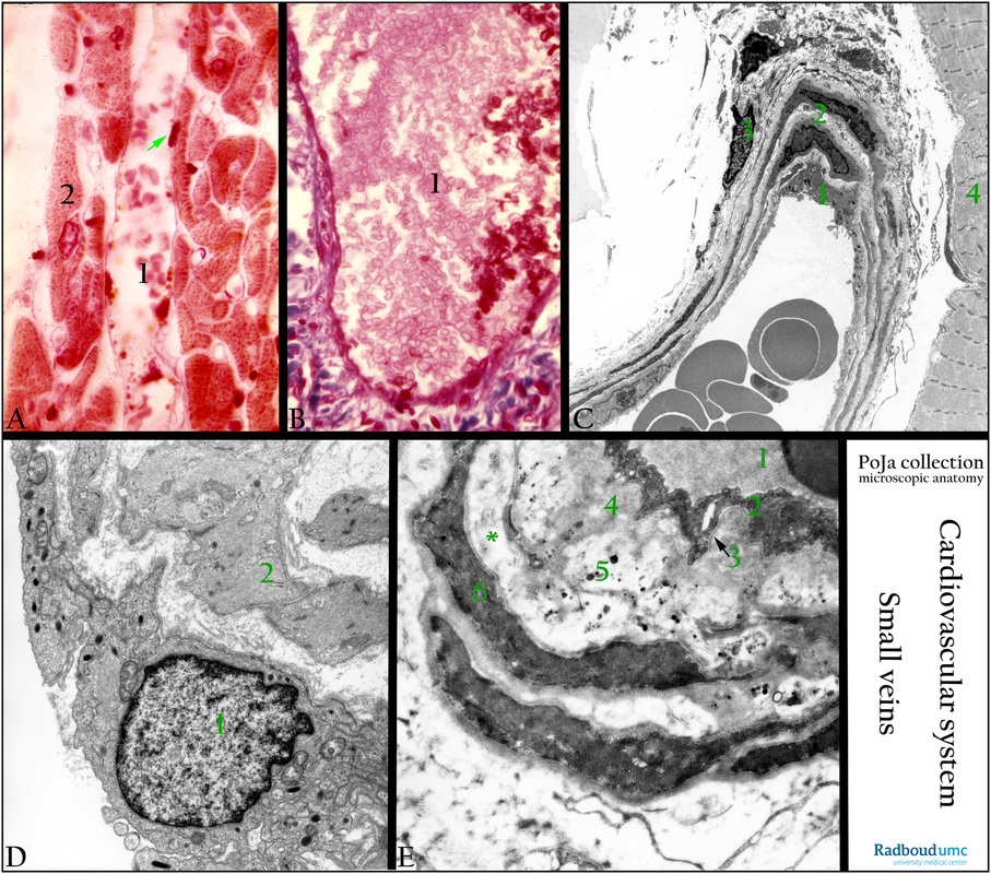
(A): Small venule (1) in heart muscle (2), Azan stain. Arrow points to the endothelial cell.
(B): Azan staining. Small vein (1) with a wide lumen in kidney. There is no internal elastic lamina (IEL) present.
(C): Electron micrograph survey small vein, with numerous pinocytotic vesicles and dense granules (Weibel-Palade bodies). (1) Endothelial cell. (2) Smooth muscle cells. (3) Fibroblast with collagen fibres. The vein is located in striated muscular tissue (4).
(D): Electron micrograph of dense-stained granules (Weibel-Palade bodies) which are found in endothelial cells of a vein. There is no internal elastic lamina between the lining endothelium (1) and the smooth muscle cells (2). The granules contain: microtubules, Willebrand factor involved in blood coagulation (vWF-polymers) , and P-selectin for recruiting passing leucocytes and several more products such as Il-8, endothelin-1, angiopoietin-2osteoprotegerin, CD63/lamp, and fucosyl-transferase VI. (Ref. 1. https://en.wikipedia.org/wiki/ Weibel%E2%80%93Palade_body). (Ref. 2. Functional architecture of Weibel-Palade bodies. Karine M. Valentijn, J. Evan Sadler, Jack A. Valentijn, Jan Voorberg, Jeroen Eikenboom. Blood May 2011, 117 (19) 5033-5043; DOI: 10.1182/blood-2010-09-267492) http://www.bloodjournal.org/content/117/19/5033?variant=long&sso-checked=true
(E): Electron micrograph small vein abdominal skin PXE patient (Pseudoxanthoma elasticum). (1) Lumen with erythrocyte, endothelium (2), accumulation of fluffy substance (4) close to the basal lamina (arrow 3). White areas represent altered elastic material (*elastin) and contain calcium crystals. Numerous electron-dense granules of varying sizes indicate calcium deposits (5), they are also found close to the smooth muscle cells (6). Pseudoxanthoma elasticum (PXE) is a disorder characterised i.a. by progressive calcification and fragmentation of elastic fibres. PXE most commonly involves elastic fibres in the reticular layer of skin, Bruch membrane in retina, and smaller/ mid-sized arteries.
Background: 1. Postcapillary venules (diameter 15-30 micrometers) drain the blood flow from the arterial capillary networks. The structure of the walls is almost identical as in arterial capillaries. The endothelium contains fenestrae and a basal lamina, however the pericytes are more branched. 2. Small pericytic venules (diameter up to 40-50 micrometers) are surrounded by pericytes. 3. Larger muscular venules (diameter 50-100 micrometers) are surrounded by pericytes and one or two layers of SMCs. Beyond the pericytes and the SMC’s a thin layer of connective tissue is present. Under the influence of e.g. histamine, serotonin, and bradykinin in inflammatory reactions the adjustable junctions of the venular endothelium ‘leak’. The results is increased permeability and local swelling. Diapedesis, the exit of leukocytes from the vasculature, occurs at this level of the microvasculature. 4. With increasing diameter loosely SMC coating of the larger muscular venules becomes more compact and are termed collecting venules (diameter 100-300 micrometers). They precede small veins (diameter 1-2 mm) followed by mid-sized veins (diameter 2-9 mm) and large veins (diameter more than 10 mm).
(partly adapted from http://medcell.med.yale.edu/histology/histology.php)
Keywords/Mesh: cardiovascular system, vascularisation, blood vessel, vein, small vein, artery, Weibel-Palade bodies, von Willebrand factor, factor VIII, calcium deposit, histology, electron microscopy, POJA collection
Title: Venous vessels: normal/ pathology (human)
Description:
(A): Small venule (1) in heart muscle (2), Azan stain. Arrow points to the endothelial cell.
(B): Azan staining. Small vein (1) with a wide lumen in kidney. There is no internal elastic lamina (IEL) present.
(C): Electron micrograph survey small vein, with numerous pinocytotic vesicles and dense granules (Weibel-Palade bodies). (1) Endothelial cell. (2) Smooth muscle cells. (3) Fibroblast with collagen fibres. The vein is located in striated muscular tissue (4).
(D): Electron micrograph of dense-stained granules (Weibel-Palade bodies) which are found in endothelial cells of a vein. There is no internal elastic lamina between the lining endothelium (1) and the smooth muscle cells (2). The granules contain: microtubules, Willebrand factor involved in blood coagulation (vWF-polymers) , and P-selectin for recruiting passing leucocytes and several more products such as Il-8, endothelin-1, angiopoietin-2osteoprotegerin, CD63/lamp, and fucosyl-transferase VI. (Ref. 1. https://en.wikipedia.org/wiki/ Weibel%E2%80%93Palade_body). (Ref. 2. Functional architecture of Weibel-Palade bodies. Karine M. Valentijn, J. Evan Sadler, Jack A. Valentijn, Jan Voorberg, Jeroen Eikenboom. Blood May 2011, 117 (19) 5033-5043; DOI: 10.1182/blood-2010-09-267492) http://www.bloodjournal.org/content/117/19/5033?variant=long&sso-checked=true
(E): Electron micrograph small vein abdominal skin PXE patient (Pseudoxanthoma elasticum). (1) Lumen with erythrocyte, endothelium (2), accumulation of fluffy substance (4) close to the basal lamina (arrow 3). White areas represent altered elastic material (*elastin) and contain calcium crystals. Numerous electron-dense granules of varying sizes indicate calcium deposits (5), they are also found close to the smooth muscle cells (6). Pseudoxanthoma elasticum (PXE) is a disorder characterised i.a. by progressive calcification and fragmentation of elastic fibres. PXE most commonly involves elastic fibres in the reticular layer of skin, Bruch membrane in retina, and smaller/ mid-sized arteries.
Background: 1. Postcapillary venules (diameter 15-30 micrometers) drain the blood flow from the arterial capillary networks. The structure of the walls is almost identical as in arterial capillaries. The endothelium contains fenestrae and a basal lamina, however the pericytes are more branched. 2. Small pericytic venules (diameter up to 40-50 micrometers) are surrounded by pericytes. 3. Larger muscular venules (diameter 50-100 micrometers) are surrounded by pericytes and one or two layers of SMCs. Beyond the pericytes and the SMC’s a thin layer of connective tissue is present. Under the influence of e.g. histamine, serotonin, and bradykinin in inflammatory reactions the adjustable junctions of the venular endothelium ‘leak’. The results is increased permeability and local swelling. Diapedesis, the exit of leukocytes from the vasculature, occurs at this level of the microvasculature. 4. With increasing diameter loosely SMC coating of the larger muscular venules becomes more compact and are termed collecting venules (diameter 100-300 micrometers). They precede small veins (diameter 1-2 mm) followed by mid-sized veins (diameter 2-9 mm) and large veins (diameter more than 10 mm).
(partly adapted from http://medcell.med.yale.edu/histology/histology.php)
Keywords/Mesh: cardiovascular system, vascularisation, blood vessel, vein, small vein, artery, Weibel-Palade bodies, von Willebrand factor, factor VIII, calcium deposit, histology, electron microscopy, POJA collection