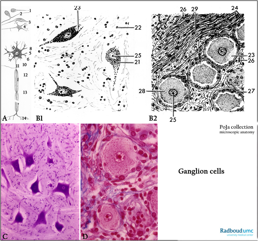
11.2.1 POJA-L3292+4433+3138+3182
Title: Comparison of intra- and extracranial neurons
Description:
(A): Scheme general structure of neuron, human.
(1) Unipolar nerve cell.
(2) Pseudounipolar nerve cell.
(3) Bipolar nerve cell.
(4) Multipolar nerve cell.
(5) Nucleus with conspicuous nucleolus.
(6) Nissl body or tigroid substance, representing ribosomes and RER.
(7) Perikaryon or cell body (cytoplasm).
(8) Dendrite.
(9) Nerves ending with “boutons terminaux” (end knobs) on the nerve cell.
(10) Axon hillock, devoid of Nissl substance.
(11) Axon or axon cylinder.
(12) Insulating myelin sheath around the axon created by the Schwann’s cell. Note its nucleus.
(13) Node of Ranvier being a myelin sheath gap occurring periodically along the axon in order to facilitate the saltatory type of conduction.
(14) Telodendron where the distal axon ramifies in small terminal branches that can end up on other nerve cells or muscle cells.
(B1): Extracranial ganglion cells: drawing of a cresyl violet-stained section of the anterior horn (cornu anterius, columna anterior), bovine. (21) Somatic motor nuclei in the anterior horn of the spinal cord, with a clear visible nucleolus (25).
(22) Nuclei of glial cells.
(23) Axon hillock devoid of Nissl substance. Note that the nerve cells are not surrounded by mantle cells or satellite cells but are embedded in a network of glial cells.
(B2): Intracranial ganglion cells of the spinal ganglion.
(23) Axon hillock.
(24) Myelinated axons.
(25) Nucleolus.
(26) Endoneurium.
(27) Mantle cells or amphicytes, i.e. neuroglial cells surrounding the neurons of ganglia. They are closely related to oligodendrocytes.
(28) Pseudo-unipolar nerve cells or peripheral ganglion cells.
(29) Nucleus of the cell of Schwann.
(C): Stain cresyl violet, bovine. Anterior column or ventral grey column with large multipolar nerve cells and nuclei of small glial cell. Equivalent to (B1).
(D): Stain Azan, bovine. Spinal ganglion cells surrounded by amphicytes, equivalent to (B2).
Keywords/Mesh: nervous tissue, spinal cord, ganglion cell, multipolar nerve cell, Nissl body, Schwann cell, amphicyte,
histology, POJA collection

