2.4 POJA-L1056
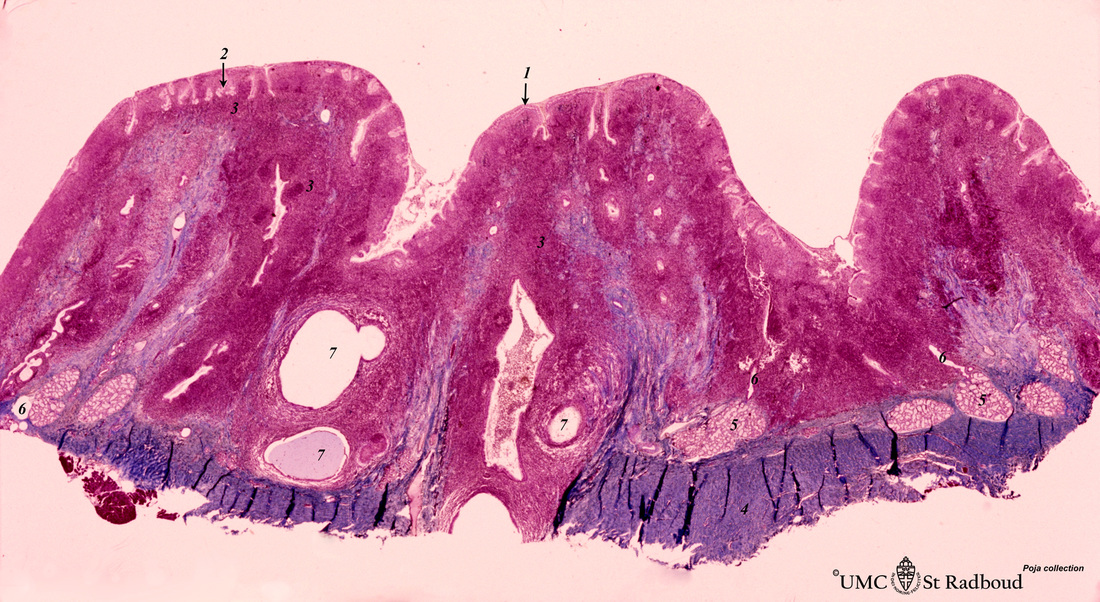
Title: Pharyngeal tonsil (‘lymphoepithelial tissues’, gut-associated lymphatic tissue or GALT) (human)
Description: Stain Azan.
The solitary pharyngeal tonsil is localized in the pharyngeal fornix and belongs to the so-called Waldeyer’s ring of pharyngeal lymphatic tissue.
A survey of this tonsil shows the folded columnar epithelium (1) with faintly light-stained goblet cells with clefts (2) in between. The lamina propria contains large aggregations of lymphatic tissue (3). The bottom wall consists of a thickened fibrous periosteum (4) of the sphenoid bone close to seromucous glands (pars nasalis pharyngis) (5). The small excretory ducts (6) of the glands open into the dilated excretory ducts (7).
Keywords/mesh: lymphatic tissue, pharyngeal tonsil, lymphoepithelial tissue, GALT, reticular tissue, histology, POJA collection

