
16.1.3 POJA-L7091+7073+7075+7094 Compact bone (cortical bone) with osteons 1
16.1.3 POJA-L7091+7073+7075+7094 Compact bone (cortical bone) with osteons 1
16.1.3 POJA-L7091+7073+7075+7094 Compact bone (cortical bone) with osteons 1
Title: Compact bone (cortical bone) with osteons 1
Description:
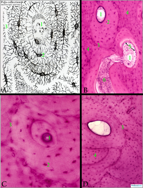
(A): Scheme of osteon in compact bone.
(B, C, D): Haematoxylin-eosin stain of human compact bone.
(1): Haversian canal system.
(2): Osteocytes.
(3): Cement line (Line von Ebner) between neighbouring osteons and interstitial lamellae (4).
(5): (B): Row of osteoblasts lining the bone of the canal system in the process of remodelling, resulting in a so-called secondary osteon.
(6): (B): Proliferation of connective tissue and blood vessels in remodelling process.
Background:
The original (foetal) fibrous or woven bone is degraded and replaced by lamellar bone (e.g., long bone, tibia). The concentric layers of compact substance have central canals (longitudinal Haversian canals) with blood vessels forming cylinders of bone tubules or osteons. The system of interconnecting transverse canal is called vascularized Volkmann’s canals (canales perforantes) being in contact with the periosteum. The remnants of degraded tubular osteons are called interstitial lamellae. The osteocytes remain embedded in their extracellular matrix with numerous thread-like extensions of cellular processes. The collagen fibres in the lamellae are arranged circular in the osteons. Different lamellae systems are separated by cement lines (ground substance with few fibrils). Modelling and remodelling appear to be age-dependent processes, higher densities of primary osteons occur between infancy and 7th years of age, but are almost completely replaced by secondary osteons after the 14th year due to remodelling. Secondary osteons are already visible after the 2nd year and reach their greatest densities in the oldest individuals.
Reference:
Cortical Histomorphometry of the Human Humerus During Ontogeny Pitfield, R., Miszkiewicz, J.J., Mahoney, P. Calcif Tissue Int 101, 148-158, 2017. DOI: 10.1007/s00223-017-0268-1
See also:
Keywords/Mesh: locomotor system, bone, compact bone, bone remodelling, osteon, lamella, Haversian canal, Volkmann’s canals, osteoblast, osteocyte, collagen, histology, POJA collection
Title: Compact bone (cortical bone) with osteons 1
Description:
(A): Scheme of osteon in compact bone.
(B, C, D): Haematoxylin-eosin stain of human compact bone.
(1): Haversian canal system.
(2): Osteocytes.
(3): Cement line (Line von Ebner) between neighbouring osteons and interstitial lamellae (4).
(5): (B): Row of osteoblasts lining the bone of the canal system in the process of remodelling, resulting in a so-called secondary osteon.
(6): (B): Proliferation of connective tissue and blood vessels in remodelling process.
Background:
The original (foetal) fibrous or woven bone is degraded and replaced by lamellar bone (e.g., long bone, tibia). The concentric layers of compact substance have central canals (longitudinal Haversian canals) with blood vessels forming cylinders of bone tubules or osteons. The system of interconnecting transverse canal is called vascularized Volkmann’s canals (canales perforantes) being in contact with the periosteum. The remnants of degraded tubular osteons are called interstitial lamellae. The osteocytes remain embedded in their extracellular matrix with numerous thread-like extensions of cellular processes. The collagen fibres in the lamellae are arranged circular in the osteons. Different lamellae systems are separated by cement lines (ground substance with few fibrils). Modelling and remodelling appear to be age-dependent processes, higher densities of primary osteons occur between infancy and 7th years of age, but are almost completely replaced by secondary osteons after the 14th year due to remodelling. Secondary osteons are already visible after the 2nd year and reach their greatest densities in the oldest individuals.
Reference:
Cortical Histomorphometry of the Human Humerus During Ontogeny Pitfield, R., Miszkiewicz, J.J., Mahoney, P. Calcif Tissue Int 101, 148-158, 2017. DOI: 10.1007/s00223-017-0268-1
See also:
- 16.1.3 POJA-L7079+7080+7082+7081 Compact bone (cortical bone) with osteons 2
- 16.1.3 POJA-L7085+7077+7078 Compact bone (cortical bone) with osteons and interstitial lamellae 3
- 16.1.3 POJA-L7083+7084 Blood vessels in compact bone (cortical bone) 4
- 16.1.3 POJA-L7112+7114+7093+7092 Remodelling of compact bone (cortical bone) 5
- 16.1.3 POJA-L7087+7088 Partially burned human bone specimen
- 16.1.3 POJA-L7090+7076 Bone: osteons in polarisation microscope
- 16.0.4 POJA-L7204 Bone: Introduction Bone formation-4
- 16.0.5 POJA-L7205 Bone: Introduction Bone formation-5 Long bones
Keywords/Mesh: locomotor system, bone, compact bone, bone remodelling, osteon, lamella, Haversian canal, Volkmann’s canals, osteoblast, osteocyte, collagen, histology, POJA collection