|
8.2 POJA-L312
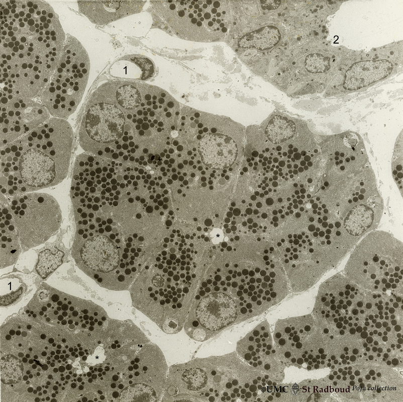
Title: Nasal glands of the nasal vestibulum (I) Description: Electron microscopy, rat. A low-power magnification reveals serous acini of a seromucous gland; in the center and lower left corner lumina (*) of the acini are present and dense secretion granules of varying sizes represent the active cells. Between the acini the distended interstitium reveals a fibroblast and two capillaries (1) At the right top corner the initial part of an intralobular draining duct (2) is seen (hardly granules!). |
|
8.2 POJA-L313
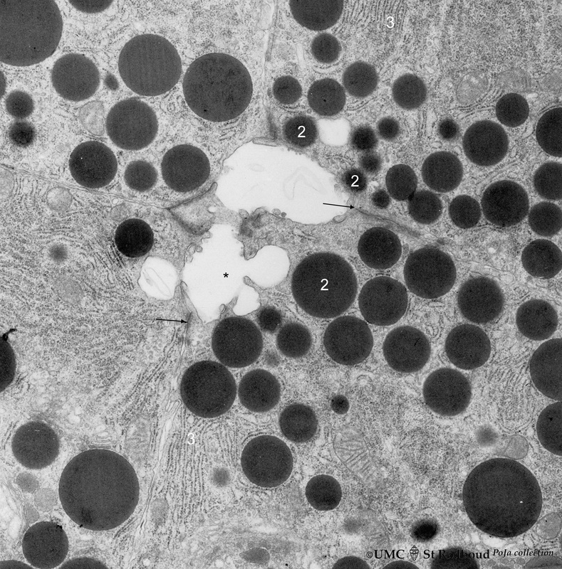
Title: Nasal glands of the nasal vestibulum (II) Description: Electron microscopy, rat. Part of a serous acinus of a seromucous nasal glands demonstrates the central lumen (*) formed by four gland cells. Note junctional complexes (↑) and the close proximity of the dense secretion granules (2) ready to be released into the central lumen. These active cells contain many secretion granules of varying sizes as well as a well-developed rough endoplasmic reticulum (3). |
|
8.2 POJA-L314
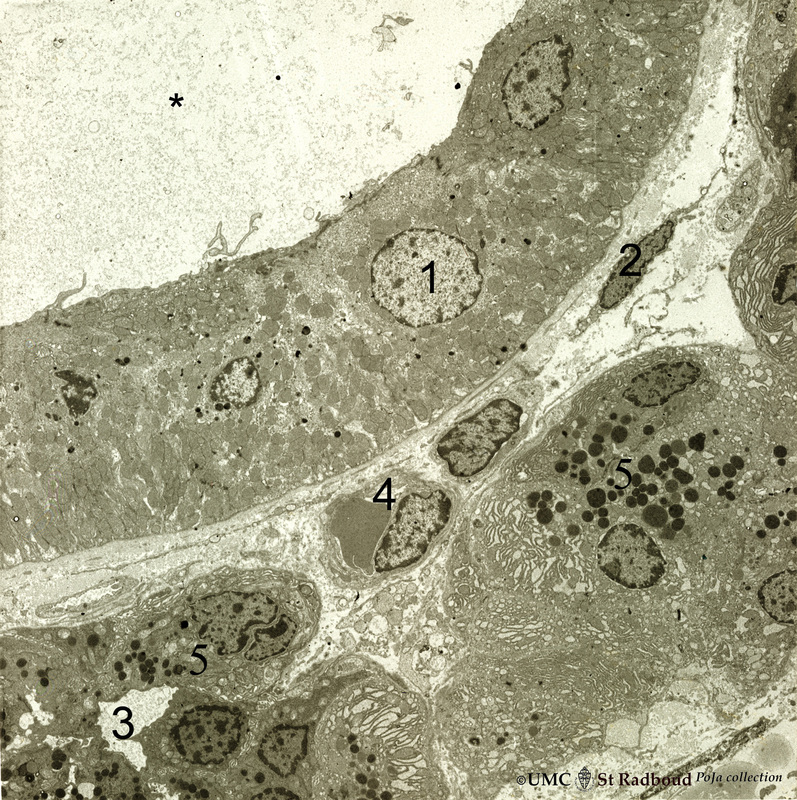
Title: Nasal glands of the nasal vestibulum (III) Description: Electron microscopy, rat. At the top part of a striated draining duct with a wide lumen (*); these cuboidal ductal cells (1) contain many basolaterally located mitochondria. The ductal cells are enforced by interstitial fibroblast (2) and a capillary (4). Neighbouring serous gland cells (5) contain dark secretion granules and widened rough endoplasmic reticulum. (3) represents lumen of the entrance of an intercalated duct. |
|
8.2 POJA-L315
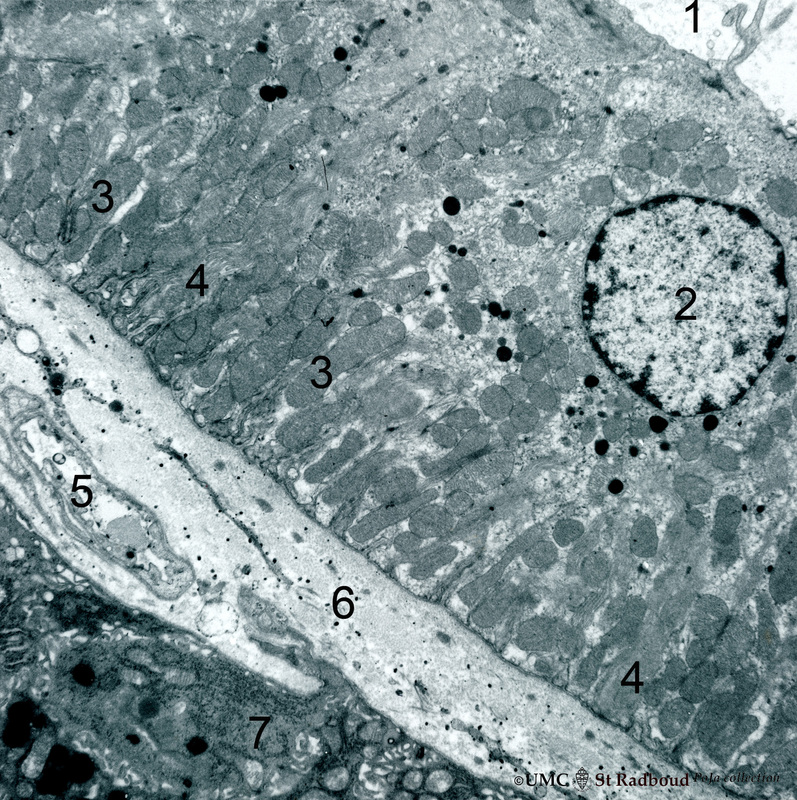
Title: Striated duct of nasal gland (IV) Description: Electron microscopy, rat. At (1) lumen of duct, (2) indicates nucleus of lining epithelial cell. Note the abundancy and palisade-like arrangement of mitochondria (3) in the basolateral regions, in the light microscope visible as a fine striation, hence the name “striated” duct or intralobular duct. (4) indicate basolateral invaginations. At (5) a capillary within a thick interstitium (6) and at (7) part of serous cell. Keywords/Mesh: respiratory tract, nasal vestibulum, nasal glands, seromucous glands, serous cells, secretion granules, rough endoplasmic reticulum, intercalated duct, striated duct, histology, electron microscopy, POJA collection |


