12.1.6 POJA-L3576+3824+2541+4399
Title: Eyelid I
Description:
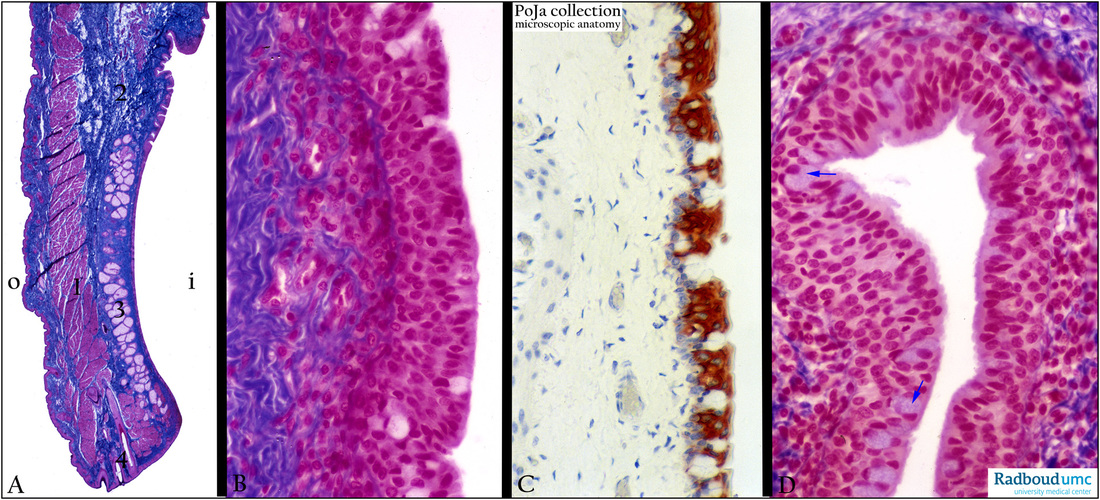
(A): Stain Azan, human.The outside part of the eyelid (O) has a skin-like structure, while the inside of the eyelid gradually displays less keratinization. The eyelid is a skin fold with a tough dense fibrous connective tissue support, the tarsal plate (blue, 2) that can be moved
by the palpebral portion of the orbicularis oculi muscle (1). At the inner side a large tree of the tarsal glands of Meibom (3) are found, holocrine sebaceous glands which open onto the tip of the eyelid. They produce an oily lipid-containing product preventing the lacrimal fluid film for drying out.
Sebaceous glands or Zeiss glands are associated with the three to four rows of hair follicles (eyelashes or cilia) (4) in the eyelid.
Apocrine sweat glands (of Moll) are non-ramifying tubular sweat glands between the hair follicles.
The conjunctiva is the mucous membrane that lines the posterior surface of the eyelids (palpebral conjunctiva) and the anterior surface of the eyeball (bulbar conjunctiva) to the sclerocorneal junction (limbus). The transition from palpebral into bulbar conjunctiva is the fornix.
(B): Stain Azan, human Detail of the palpebral conjunctival epithelium close to the fornix with multilayered (4-5) cylindrical epithelial cells including goblet cells.
(C): Immunoperoxidase staining with DAB and anti- keratin monoclonal antibody RCK102, rat.
Palpebral conjunctival epithelium: the immunoperoxidase staining with RCK102 (cytokeratin 5 and 8) stains predominantly the intermediate and superficial cells but not the basal cells and the goblet cells in this epithelium, which is thinner than in human.
(D): Stain Azan, fornix, human. Note many goblet cell types (arrows) especially in the fornix.
Keywords/Mesh: eye, eyelid, conjunctiva, fornix, tarsal gland, meibomian gland, Zeiss gland, Moll gland, histology, POJA collection

